
Page 69 - 2019_11 Resto del Mondo-web
P. 69
Loss of Notch1 TAD interferes with niche recovery
and endomucin+ microvessels and a high frequency of apoptotic EC. Defective regeneration of EC in these mice caused gaps in sinusoidal vessels leading to severe hemor- rhage.5,50 The occupancy of the Notch transcriptional com- plex and expression of Notch target genes associated with survival and proliferation, such as Hes1 and Myc,51,52 was markedly decreased in Notch1+/ΔTAD BM EC, indicating that
the regeneration of niche EC following chemotherapy was dependent on high levels of Notch signaling. Validation was obtained using conditional deletion of the Notch1 receptor in the VE-cadherin+ EC. Similar to the Notch1+/ΔTAD, this EC- specific, conditional, loss-of-function Notch1 model exhib- ited defective regeneration of BM EC highlighted by increased EC apoptosis and limited hematopoietic recovery.
AB
CD
EF
G
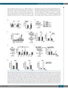
Figure 7. Notch1 functions downstream of Tie2 signaling to mediate endothelial niche recovery. (A) Representative gating strategy for osteoblasts (Ter119-CD45- CD31-Sca-1-CD51+ cells) in digested bones from mice treated with phosphate-buffered saline (PBS) and 5-fluorouracil (5-FU) (PBS, n=5; 5-FU, n=15; left panel). Expression of Ang1 was determined by quantitative reverse transcriptase polymerase chain reaction (RT-qPCR) in osteoblasts and Lin-Sca-1+ cells treated with PBS and 5-FU (right panel). GAPDH was used as an internal expression control. (B) Schematic of Ang1 treatment in serum-starved (16 h) cultured bone marrow-derived endothelial cells (cBEC) (left panel). Expression of p-Tie2, cleaved Notch1 and GAPDH protein levels in Ang1-treated cBEC was detected by western blot (right panel). (C) RT-qPCR expression of Notch target genes from cBEC treated as described in (B). Values are normalized to those in PBS-treated cBEC. GAPDH was used as an internal expression control. (D) Schematic of Tie2 inhibitor (Tie2i) and Ang1 treatment in serum-starved (16 hr) cBEC (left panel). Expression of Dll4 and Jag1 was determined by RT-qPCR in Tie2i and/or Ang1-treated cBEC. The vehicle for the Tie2 inhibitor was dimethysulfoxide (DMSO). Vehicle for Ang1 is PBS. (E) Schematic for treatment of cBEC with γ secretase inhibitors (GSI). RT-qPCR expression analysis of the indicated genes in cBEC treated with GSI/washout. Values were normal- ized to those of DMSO-treated cBEC. GAPDH was used as an internal expression control. (F) Schematic for treatment of control PMIGR1 or PMIGR1-ICN∆TAD cBEC with 5-FU and Ang1. The indicated cBEC populations of cells were monitored for growth after 5-FU and Ang1 treatment. (G) RT-qPCR expression of Tie2 target Socs3, and Notch targets Hes1 and Hey1 was examined after cell growth analysis of cBEC in (F). Values were normalized to those of PMIGR1 cBEC without Ang1-treatment. GAPDH was used as an internal expression control. Unless indicated differently, n=3 were used for experiments in this figure. *P<0.05, **P<0.01, ***P<0.001.
haematologica | 2019; 104(11)
2175
