
Page 157 - 2019_11 Resto del Mondo-web
P. 157
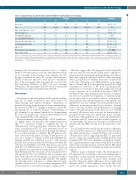
Identifying Richter’s after BCRi Therapy
Table 4. Characteristics of patients who underwent Richter’s transformation on venetoclax.
Patients 123456
Summary
64 (56 – 66)
3 (50) 12 (6 – 20) 5.5 (2 – 9) 4 (67)
Age,† years
Sex‡
Time from diagnosis,† years Prior therapies,† n del(17p)/TP53 mutation‡ Screening PET-CT SUVmax† Largest node at baseline,† cm β-2 microglobulin,† mg/L ALC, x109/L†
Best response on venetoclax
Time to RT,† weeks
56 65
Male Female 12 12
5 9 Yes Yes 11 8 4.8 10.8 2.5 5.7 20 2 PR SD
30 19
58 63
Female Male 9 6 9 2 Yes No 5 <3 2.5 7.2 2.8 6.8 71 6 PR PR 87 68
65 66
Female Male 18 20 6 5 No Yes
6 <3 7(<3–11)
3.1 8.6 N/A 2.8 406 .9 CR* PR 72 71
4.8 (2.5 – 10.8) 2.8 (2.5 – 6.8) 13 (0.9 – 406) 83% ORR 69.5 (19 – 87)
del(17p): chromosome 17p deletion; PET-CT: positron emission tomography-computed tomography; SUVmax: maximum standardized uptake value of 18-F-fluorodeoxyglucose; N/A: data not available; ALC: absolute lymphocyte count; PR: partial remission; SD: stable disease; CR: complete remission; ORR: overall response rate (CR+PR); RT: Richter’s transformation.*Patient had PR at weeks 8 and 24 and then CR at week 60.†Summary column describes the median (range) across the six patients.‡Summary column describes the number (n) (%) across the six patients.
patients who discontinued venetoclax due to a relapse with RT, two met criteria for biopsy and underwent biop- sy at screening. Both biopsies were negative for RT, though one patient had prior RT and the other had prior DLBCL both more than two years prior to enrollment. Five of the six patients who developed RT on venetoclax had screening SUVmax on PET <10 and screening SUVmax for all six patients with RT was not statistically different from other enrolled patients (P=0.24).
Discussion
The diagnosis and management of RT remain challeng- ing due to rapid progression and refractoriness to chemotherapy and targeted therapies.3 Evaluation of patients following chemotherapy or chemoimmunothera- py showed that PET-CT imaging is helpful to detect RT and identify sites to target for biopsy,3,9,10 with SUVmax of ≥10 identified as an optimal threshold for distinguishing RT versus CLL progression,10 and associated with poor sur- vival, independently of RT diagnosis.3
Newer treatments, including BCRi or BCL-2 inhibitors, are highly active for patients with relapsed/refractory CLL15,16,21,22 and have begun to supplant the use of chemotherapy. Data reported here represent the largest series of prospective PET-CT scans performed based on predetermined criteria in patients following discontinua- tion of either ibrutinib or idelalisib. Of 167 patients who were screened for this phase II study, 57 met protocol cri- teria for a biopsy to evaluate for RT following PET-CT imaging, and RT was confirmed for 4.8% of all screened patients. Five of these patients had SUVmax ≥10 while the other two had SUVmax <10 with other factors associated with RT (e.g. LDH elevation or B symptoms). Based on this analysis, we report the test characteristics for PET-CT with SUVmax ≥10. We observed a lower sensitivity (71%) and specificity (50%) for SUVmax ≥10 in patients who have been exposed to BCRi therapy as compared to prior reports in patients not progressing on BCRi.10 We empha- size the importance of biopsy to confirm clinical or radi- ographic suspicion of RT or a secondary malignancy. PET- CT should not be used without biopsy to diagnose RT.
Our data suggest that CLL progression following BCRi exposure may be more metabolically active compared to progression following chemoimmunotherapy. In addition, these data suggest no difference in FDG avidity in patients with CLL regardless of whether they were on their BCRi at the time of imaging or not. Only LDH above the upper limit of normal and β-2 microglobulin ≥3 mg/L had high sensitivity for detection of RT from CLL progression. Limitations of our study are the small sample size in each group of patients on or off BCRi at the time of PET-CT and the fact that other patient features (such as cytogenet- ics) were not available for all patients, so statistical analy- ses for those factors only include subsets of the patients with available data. Another limitation of the analysis was that PET-CT was not centrally reviewed and there may be differences in imaging methodologies, number of lymph nodes assessed per study, and interpretations of imaging studies. Though pathology results were also not centrally reviewed, all biopsies were reviewed by hematopatholo- gy experts at the study sites, which were major academic institutions. Although there was a relatively small number of patients with RT events included in these analyses, to our knowledge, this dataset is the largest of BCRi-treated patients who had PET-CT and were prospectively evalu- ated.
For 127 patients who enrolled in the study, no difference was seen in the response rate with venetoclax monother- apy when stratified by screening SUVmax of 10 in whom a biopsy ruled out RT, suggesting that PET-CT performed prior to venetoclax initiation may identify patients with an inferior PFS. We have identified a population of high- risk CLL patients previously treated with BCRi for whom venetoclax monotherapy may result in inferior PFS and in whom venetoclax-based combination regimens should be studied.
Whereas in prior ibrutinib studies, the development of RT appeared to be a relatively early event (median approx. 6 months),23 in this study, the development of RT on vene- toclax for six patients occurred later (median 12.8 months). This may reflect differences in how individual therapies (BCRi vs. BCL2 inhibitor) affect the CLL clone and/or the patient populations. More likely, the later
haematologica | 2019; 104(11)
2263
