
Page 156 - 2019_11 Resto del Mondo-web
P. 156
A.R. Mato et al.
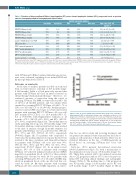
Table 3. Detection of biopsy-confirmed Richter’s transformation (RT) versus chronic lymphocytic leukemia (CLL) progression based on positron emission tomography-computed tomography and clinical factors.
PET-CT SUVmax <10/≥10 PET-CT SUVmax <5/≥5
PET-CT SUVmax <11/≥11 PET-CT SUVmax <12/≥12 Lactate dehydrogenase ≤/>ULN SPD at baseline
TP53* mutated/unmutated IGHV * mutated/unmutated CD-38* positive/negative ZAP-70* positive/negative
β2 microglobulin * </≥3 mg/L
Sensitivity Specificity PPV NPV
71% 50% 26% 88%
71% 4% 16% 33%
71% 61% 31% 89%
57% 68% 31% 86%
83% 29% 20% 89%
67% 48% 24% 86%
60% 63% 25% 88%
25% 86% 25% 86%
25% 40% 6% 77%
50% 23% 9% 75%
75% 25% 20% 80%
ROC area
61%
63%
66%
63%
56%
59%
61%
56%
68%
64%
50%
Odds ratio [95% CI], logistic P
2.5 [0.4–15], P=0.318 0.09 [0.01–1.2], P=0.071 3.8 [0.6–24], P=0.143 2.8 [0.5–15], P=0.231 2 [0.2–20], P=0.554
1 [1–1], P=0.244
2.5 [0.35–18], P=0.362 2.1 [0.16–28], P=0.569 0.22 [0.02–2.5], P=0.220 0.3 [0.01–6.4], P=0.440 1 [0.08–13], P=1.0
Logistic regression analyses
PPV: positive predictive value; NPV: negative predictive value; ROC: Receiver Operator Characteristic; PET-CT: positron emission tomography-computed tomography; SUVmax: maximum standardized uptake value of 18-F-fluorodeoxyglucose; ULN: upper limit of the normal range; SPD: sum products of the greatest transverse diameters (tumor size). *Site-reported data.The logistic regression analyses only included patients with available data.
with SUVmax ≥10. Other baseline clinical/prognostic fea- tures were evaluated, including above normal LDH and β2-microglobulin levels (Table 3).
Outcomes on venetoclax
At the time of analysis, patients enrolled on study had been on venetoclax for a median of 10.3 months (range: 0.1-26 months). Falchi et al. had previously reported that patients with SUVmax ≥10 had an inferior survival on chemotherapy/chemoimmunotherapy;3 therefore, we evaluated outcomes of venetoclax using this cutoff of screening SUVmax. The ORR on venetoclax was 65% (82 of 127) for all enrolled patients, and was similar when stratified by screening PET-CT SUVmax <10 (65%, 74 of 114) versus ≥10 (62%, 8 of 13) (P=0.81). Median progres- sion-free survival (PFS) was longer for venetoclax-treated patients with screening PET-CT SUVmax <10 versus ≥10 [24.7 months (95%CI: 20.1, -) vs. 15.4 months [95% CI: 0.4, -]; P=0.0335), with Kaplan-Meier estimates at 12 months of 79% (95%CI: 69%, 85%) and 58% (95%CI: 27%, 80%), respectively (Figure 1A). The median time on study, including follow up, was 13.8 months (range: 0.03- 31 months). Median overall survival had not been reached at the time of analysis, though 12-month estimates for patients with SUVmax <10 versus ≥10 were 94% (95%CI: 87%, 97%) and 76% (95%CI: 43, 92%), respectively (P=0.061) (Figure 1B). For patients who responded to venetoclax, the median duration of response had not been reached at the time of analysis, and 12-month estimates for patients with SUVmax <10 versus ≥10 were 90% (95%CI: 80%, 96%) and 75% (95% CI: 32%, 93%), respectively (P=0.17) (Figure 1C). In an intent-to-treat analysis, 29% (33 of 114) of patients with screening SUVmax <10 achieved minimal residual disease (MRD) negativity in peripheral blood, with 19 of these patients achieving this outcome by week 24. For patients with screening SUVmax ≥10, 23% (3 of 13) achieved MRD neg- ativity in peripheral blood, with one who had this out- come by week 24. Of three patients with screening SUVmax ≥10 who achieved MRD negativity on veneto-
Figure 2. Time to chronic lymphocytic leukemia (CLL) progression or Richter’s transformation (RT) on venetoclax. Shown is the cumulative incidence of CLL progression or Richter’s transformation on venetoclax. Thirty-three patients dis- continued venetoclax due to CLL progression and six due to biopsy-confirmed RT following both imaging and clinical changes. Median time to CLL progression was 8.5 months (range: 0.1-28 months) and to RT was 12.8 months (range: 4.4–19.7 months). Tick marks represent patients with events.
clax, two are still on study and receiving treatment, and one patient discontinued venetoclax and later died due to squamous cell carcinoma of head and neck. There was no statistical difference in the rate of MRD-negativity in blood based on screening SUVmax ≥10 versus <10 (P=0.83).
Sixty-two (49%) patients discontinued venetoclax (Online Supplementary Figure S1), with a median time to discontinuation for any reason of 8.6 months (range: 0.1- 28). Thirty-three patients (26%) discontinued venetoclax due to CLL progression and six (4.7%) due to biopsy-con- firmed RT following clinical findings and radiographic changes (Table 4). The median time to CLL progression was 8.5 months (range: 0.1-28 months) and to RT was 12.8 months (range: 4.4-19.7 months) (Figure 2). Of six
2262
haematologica | 2019; 104(11)
