
Page 122 - 2019_11 Resto del Mondo-web
P. 122
X. Li et al.
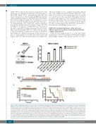
CUDC-907 for 24 h caused an increase in annexin V+ cells, which was accompanied by increased cleaved caspase 3 and PARP (Figure 1B-D), demonstrating that the cells underwent apoptosis. Short hairpin (sh)RNA knockdown of Bax and Bak partially rescued U937 cells from CUDC- 907-induced apoptosis (Figure 1E). Furthermore, overex- pression of Bcl-xL abolished CUDC-907-induced apopto- sis demonstrating that CUDC-907 induces apoptosis through the intrinsic apoptosis pathway (Figure 1F). The potential in vivo efficacy of CUDC-907 was evaluated in an early stage MV4-11-derived xenograft mouse model. Mice were treated with CUDC-907 daily for 8 days, given 4 days off treatment, and then treated daily for another 6 days (Figure 1G). All mice were given a 4-day break due to the 3% body weight loss in the mice treated with 150 mg/kg CUDC-907 after the initial eight doses (Figure 1H).
F
This body weight loss was completely reversible within 4 days. The median survival following CUDC-907 treat- ment was 44 days for the animals given the 100 mg/kg dose and 47 days for those given the 150 mg/kg, which are 11 and 14 days longer (or 33.3% and 42.2% increases in lifespan), respectively, than the median survival of the mice given the vehicle control (33 days; P=0.002) (Figure 1I). These results suggest that CUDC-907 treatment pos- sesses modest antileukemic activity in vivo.
CUDC-907 treatment decreases viable cells and induces apoptosis in primary acute myeloid leukemia samples from patients
CUDC-907 IC50 ranged from 8.1 to 1,831 nM (Online Supplementary Table S2) in primary AML patient samples. Interestingly, samples from patients positive for FLT3-
G
HI
Figure 1. Figure 1. CUDC-907 treatment decreases viable cells and induces apoptosis in acute myeloid leukemia cell lines and shows promise in an acute myeloid leukemia cell line-derived mouse model. (continued from the previous page) (F) MV4-11 cells were infected with Precision LentiORF Bcl-xL and RFP control overex- pression lentivirus particles overnight, then washed and incubated for 48 h prior to addition of blasticidin to the culture medium. Whole cell lysates were subjected to western blotting. The fold changes for the Bcl-xL densitometry measurements, normalized to β-actin and then compared to no drug treatment control, are indi- cated (left panel). The cells were treated with CUDC-907 for 24 h and then subjected to annexin V/PI staining and flow cytometry analysis. ***P<0.001 (right panel). (G-I) MV4-11 cells (1 x 10⁶ cells/mouse) were injected through the tail vein of immunocompromised NSGS mice. Three days after cell injection the mice were ran- domized (5 mice/group) and treated with vehicle control (3% 200 proof ethanol, 1% polyoxyethylene 20 sorbitan monooleate, and USP water), 100 mg/kg CUDC- 907, or 150 mg/kg CUDC-907 for 8 consecutive days followed by 4 days off treatment, and then an additional 6 days of treatment. (H) Body weights were measured on a daily basis and are shown as mean ± SEM. (I) Overall survival probability, estimated with the Kaplan-Meier method. AML: acute myeloid leukemia; CUDC: CUDC- 907; NTC: non-treated control; RFP: red fluorescent protein; cf-Caspase 3: cleaved caspase 3; cf-PARP: cleaved PARP.
2228
haematologica | 2019; 104(11)
