
Page 172 - 2019_10 resto del Mondo_web
P. 172
V.G. Ramakrishnan et al.
(T163) is a well-documented post-translational modifica- tion that stabilizes MCL-1, protecting it from proteasomal degradation.28 However, we did not observe a correlation between p-MCL-1 at serine 64 (S64) and BIM binding preference (data not shown). p-MCL-1 (S64) increases the binding capacity of MCL-1, but not its stability.29
To our surprise, p-BCL-2 (S70) and p-MCL-1 (T163) were nearly perfect in predicting sensitivity to either MEK + HDAC or BCL-2 + HDAC inhibition (Figure 5A). This was particularly striking in the case of DOX40, a doxoru- bicin-resistant cell line derived from RPMI8226. DOX40 expressed more p-BCL-2 (S70) than p-MCL-1 (T163), and was sensitive to the ABT-199/LBH589 combination, whereas its parental cell line RPMI8226 did not express p- BCL-2 (S70), but did express p-MCL-1 (T163), and was
sensitive to the AZD6244/LBH589 combination (Figure 5A). Interestingly, the only cell line we tested that was resistant to both drug combinations was KMS11, and this line has low expression of both p-BCL-2 (S70) and p- MCL-1 (T163) (Figure 5A).
RAS/RAF mutational status predicts sensitivity to MEK + HDAC or BCL-2 + HDAC inhibition
Since MEK inhibitors target the pathway downstream of RAS/RAF, we were curious to determine whether sen- sitivity to the AZD6244/LBH589 combination correlates with RAS/RAF mutational status. This is a felicitous prospect because the MEK/ERK pathway in part controls p-MCL-1 (T163).28 Indeed, the RAS-mutated cell lines H929, MM1R, MM1S, RPMI8226, and the RAF-mutated
AB
CE
D
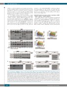
Figure 4. Predominance of BIM:MCL1 complexes correlates with sensitivity to MEK + histone deacetylase (HDAC) inhibition; MEK + HDAC inhibitor-resistant cell lines that had predominantly BIM:BCL-2 complexes were synergistically killed by BCL-2 + HDAC inhibition. (A) Whole-cell lysates from a panel of human multiple myeloma (MM) cell lines were immunoprecipitated with BIM. Subsequently, western blotting was performed to examine baseline levels of BIM:BCL-2, BIM:BCL-XL and BIM:MCL-1 complexes. Whole cell lysates (input) were also separated with sodium dodecylsulfate polyacrylamide gel electrophoresis (SDS-PAGE) and probed for the indicated proteins. Cell lines sensitive to the AZD6244/LBH589 combination are indicated in blue. The mutational status of the cell lines, i.e. mutated or wild-type (WT) for RAS/RAF, is shown. (B) The BH3 mimetic ABT-199 and the pan-HDAC inhibitor LBH589 induced synergistic cytotoxicity (assessed using MTT) by 72 h in the human MM cell lines KMS18, OPM2 (RAS/RAF wild-type) and KMS28 (RAS mutant). Viability is shown as percent of control on the Y-axis. Combination index (CI) val- ues <1.0, indicating synergy, are shown for each cell line. (C) KMS28 and OPM2 were treated with ABT-199/LBH589 for 24 h, then whole-cell lysates were separated using SDS-PAGE and probed for the indicated proteins. (D) OPM2 was treated with 50 nM ABT-199 and 5 nM LBH589 for 24 h, then immunoprecipitates for BCL-2 and MCL-1, and whole cell lysates (input) were separated using SDS-PAGE and probed for the indicated proteins. (E) OPM2 and KMS28 were treated with ABT-199 (50 nM and 250 nM, respectively) and LBH589 (5 nM) for 12 h. BAX and BAK immunoprecipitates and whole-cell lysates (input) were separated using SDS-PAGE and subjected to western blotting for the indicated proteins. All experiments were performed in triplicate.
2068
haematologica | 2019; 104(10)
