
Page 171 - 2019_10 resto del Mondo_web
P. 171
HDAC + MEK or BCL-2 inhibition in multiple myeloma
Finally, we noted enhanced apoptosis after treating plas- ma cells from MM patients with the ABT-199/LBH589 combination (Table 1).
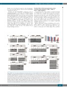
Notably, the ABT-199/LBH589 combination did not synergistically induce cell death in the RAS/RAF mutant, AZD6244/LBH589-sensitive cell lines (i.e. H929, MM1R, MM1S, RPMI and U266). This dovetails with the finding that while LBH589 dissociated BIM:MCL-1 complexes in all the cell lines we tested, it did not shift BIM onto BCL- 2 in RAS/RAF mutant cell lines (e.g. MM1S) (Figure 3C). However, LBH589 did shift BIM onto BCL-2 in the RAS/RAF wild-type cell lines (e.g. OPM2) (Figure 4D), which jibes with its potent synergistic effect when com- bined with ABT-199. In summary, it appears that AZD6244/LBH589 and ABT-199/LBH589 target two dis- tinct subgroups of MM cell lines with different BCL-2 family binding proclivities.
AB
CD
Baseline MCL-1/BCL-2 phosphorylation status correlates with sensitivity to MEK + HDAC or BCL-2 + HDAC inhibition
To determine why certain cell lines would preferentially have BIM bound to one anti-apoptotic protein over anoth- er, i.e. BCL-2 versus MCL-1, we examined several phos- phorylation sites known to affect the binding capacity and stability of the BCL-2 family to see if there was any corre- lation. Interestingly, cell lines with higher p-BCL-2 at ser- ine 70 (S70) tended to have more BIM bound to BCL-2 (Figure 5A). p-BCL-2 (S70) is known to increase the anti- apoptotic capacity of BCL-2, i.e. its ability to bind BAK and BH3-only proteins.27
On the other hand, cell lines with BIM mostly bound to MCL-1 tended to have relatively low expression of p- BCL-2 (S70), as well as relatively high expression of p- MCL-1 at threonine 163 (T163) (Figure 5A). p-MCL-1
Figure 3. MEK + histone deacetylase inhibitor induced synergistic apoptosis is mediated by BIM. (A) The RAS mutant human multiple myeloma (MM) cell lines H929 and MM1S were treated with AZD6244/LBH589 for 24 h, then whole-cell lysates were blotted for the indicated proteins. (B) MM1S was electroporated with scram- bled siRNA or BIM siRNA and then left untreated or treated with 5 nM LBH589. At 72 h, cell viability was assessed using flow cytometry by analyzing the proportion of annexin–/propidium iodide (PI)– cells, shown as percent of control on the Y-axis. Furthermore, the whole-cell lysates were separated using sodium dodecylsulfate polyacrylamide gel electrophoresis (SDS-PAGE) and subjected to western blotting for the indicated proteins to confirm silencing. Error bars represent the standard error of mean of triplicate experiments. Differences between groups were calculated with the Student t test. **P<0.001. (C) (Upper) H929 and MM1S were treated with AZD6244 (250 nM and 150 nM, respectively) and LBH589 (5 nM) for 24 h. BIM immunoprecipitates were separated using SDS-PAGE and subjected to western blotting to examine BCL-2, MCL-1 and BCL-XL binding patterns. Whole cell lysates (input) were also separated and probed for the indicated proteins. (Lower) Immunoprecipitates from MM1S for BCL-2 and MCL-1 were also separated and probed to examine BIM binding. (D) H929 and MM1S were treated with AZD6244 (250 nM and 150 nM, respectively) and LBH589 (5 nM). BAX and BAK immunoprecipitation was performed and western blotting was used to examine levels of BIM and MCL-1 bound to BAX and BAK. Whole cell lysates (input) were also separated and probed for the indicated proteins. All experiments were performed in triplicate.
haematologica | 2019; 104(10)
2067
