
Page 126 - 2019_10 resto del Mondo_web
P. 126
P.-Y. Dumas et al.
ABC
DE
FG
H
I
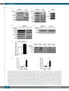
Figure 3. Activation of STAT5 up-regulates AXL gene expression and phosphorylation. (A) UT7-mpl acute myeloid leukemia (AML) cells were treated with GM-CSF (2.5 ng/mL) or TPO (20 nM) for the indicated time and lysed. Immunoblot analysis of the indicated proteins; HSP60 was used as a loading control. (B) UT7-mpl cells were treated with GM-CSF (2.5 ng/mL) or TPO (20 nM) for the indicated time in the absence or presence of pimozide (pi, 2 μM), JAK inhibitor-I (ji, 1 μM) or Ly294002 (ly, 20 μM). Total cell lysates were analyzed by western blotting with the indicated antibodies. HSP60 was used as a loading control. (C) UT7-mpl cells were incubated with (+) or without (-) TPO (20 nM) for one hour (h) before being lysed. AXL immunoprecipitates were prepared and analyzed by immunoblotting with the indicated antibodies. (D and E) MOLM-14 or MV4-11 cells were pre-incubated with (+) or without (-) AC220 (3 nM) for 3 h before adding IL-3 (ng/mL, MOLM-14) or TPO (nM, MV4-11) at the indicated concentration. Cells were incubated for 4 h before being lysed. (F and G) MS5 co-cultured AML primary blasts were maintained in serum-free medium without stroma for 18 h before being incubated in the presence of a IL-3 (20 ng/mL)/GM-CSF (10 ng/mL)/TPO (20 nM) cytokine cocktail for 7 h and then lysed for protein (F, n=6, AML#10-12 and 15-16) and mRNA (G, n=5, AML#10-14) purification. Immunoblot analysis of the indicated proteins using β actin or HSP60 as loading controls; real-time quantitative polymerase chain reaction (RT-qPCR) quantification of AXL mRNA levels in cytokine-treated and untreated primary AML cells, normalized to GAPDH expression, and expressed relative to untreated cells. (H and I) UT7-mpl cells were incubated with (+) or without (-) TPO (20 nM) for 30 minutes. Chromatin- immunoprecipitation (IP) assays were performed using control immunoglobulins (IP IgG), STAT5 (IP STAT5) or RNA polymerase II (IP PolII) antibodies. Immunoprecipitated DNA were analyzed by qPCR using primers spanning the conserved STAT5-responsive element of AXL gene sequence and expressed relative to total lysates (input). Graphs show the mean±Standard Error of Mean of at least three independent experiments. *P<0.05; **P<0.01; ***P<0.001; NS: not significant.
2022
haematologica | 2019; 104(10)
