
Page 128 - 2019_10 resto del Mondo_web
P. 128
P.-Y. Dumas et al.
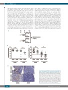
engraftment (timing, intensity) between MV4-11 shCtrl and MV4-11 shAXL cells after seven days (Online Supplementary Figure S5B). Half of the mouse cohort was then treated with AC220 (5 mg/kg/day) by daily oral gav- age for seven days, and response to treatment was assessed by BLI at day 14. The tolerance to treatment was checked by body weight control twice a week. A similar bioluminescence indicative of tumor progression was observed for MV4-11 shCtrl and MV4-11 shAXL cells in the vehicle-treated cohort (Figure 5A and B). In contrast, a significant decrease in total bioluminescent signal was observed in response to AC220 in MV4-11 shAXL- engrafted mice as compared to MV4-11 shCtrl-engrafted animals (P<0.01) (Figure 5B). To strengthen these data in the specific setting of the microenvironment, another set of experiments was performed with the same sequence of conditioning, engraftment and treatment, but BLI was per- formed upon treatment on femurs and tibias ex vivo at sac-
rifice. Again, a significant decrease in bioluminescence was observed in bones from AC220-treated MV4-11 shAXL-engrafted mice compared to bones from MV4-11 shCtrl-engrafted animals, whereas no significant differ- ence was observed between these two cohorts in the absence of treatment (Figure 5C). Immunohistochemistry analysis of BM was then performed to trace human cells in the murine bone marrow tissue. Similar amounts of human cells were detected in MV4-11 shCtrl- and shAXL- injected mice in the absence of treatment (Figure 5D). In the AC220-treated cohort, as expected, lower amounts of human cells were detected in MV4-11 shCtrl-engrafted BM than in the untreated cohort. Interestingly, almost no human cells were detectable in MV4-11 shAXL-treated animal samples, so BLI detection was under the threshold in MV4-11 shAXL-treated animals (Figure 5D). Altogether, these results show that AXL plays a key role in the AC220 response of FLT3-ITD AML cells in the specific context of
B
D
A
Figure 5. AXL sustains FLT3-ITD acute myeloid leukemia (AML) cell resistance to AC220 in the bone marrow hematopoietic niche. (A) MV4-11 shCtrl-Luc and MV4-11 shAXL-Luc were injected at day 0 (106 cells/mouse) in the retro-orbital sinus vein of busulfan-pre- treated NSG female mice. Mice were then treated or not with AC220 (5 mg/kg/day body weight) from day 8 to day 14. At day 14, animals (B) or bones (femurs, tibias), collected from the indicated animals (C) were subjected to bioluminescent imaging (BLI). The dotted line shows the median background signal indicating the threshold for BLI sensitivity. Ph: photon; s: second; sr: steradian. (D) Immunohistochemistry analysis of Human Leukocyte Antigen expression in bone marrow biopsies collected from the indicated AML cell-injected mice in the absence or presence of AC220 at day 14. *P<0.05; **P<0.01; ***P<0.001; NS: not significant.
2024
haematologica | 2019; 104(10)
C
