
Page 125 - 2019_10 resto del Mondo_web
P. 125
AXL and resistance to quizartinib in the hematopoietic niche
transcription factors bind to TTCN3GAA STAT-Response Element (SRE) on their target genes. One SRE that is con- served among species including rat, mouse and human was identified within intron 16 and located at Chr19(+) [41,725,104-41,767,672] (www.genome.ucsf.edu). Oligonucleotide pull-down assays showed that this iden- tified SRE was functional, since it bound STAT5 as effi-
AB
C
ciently as the control canonical SRE (ONc) from the IRF1 promoter sequence (Online Supplementary Figure S3B). Chromatin immunoprecipitation (ChIP) assays with STAT5 antibodies showed that STAT5-activation trig- gered binding to the AXL genomic sequence (Figure 3H). Together with STAT5 binding, recruitment of RNAPolII to the AXL gene was enhanced, as assessed by anti-RNAPolII
DE
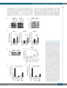
Figure 2. STAT5 up-regulates AXL which contributes to FLT3-ITD acute myeloid leukemia (AML) cell survival. (A) MOLM-14 and MV4-11 cells were transduced with the indicated shRNA encoding lentiviral vectors and lysed three days later. Immunoblot analysis of the indicated proteins was per- formed with β actin as a loading control. (B) MOLM-14 and MV4-11 cells were co-cul- tured in the absence (-) or presence (+) of OP9 stromal cells for 48 hours (h) before being isolated and lysed. The indicated pro- teins were analyzed by immunoblotting. (C) MV4-11, MOLM-13 and MOLM-14 cells were treated in the absence (Vehicle) or presence of minimal effective (MV4-11, MOLM-14) or suboptimal (MOLM-13) dose of AC220 (1 nM) and R428 (0.3 μM) or both (Combo) for 48 h. Apoptosis induction was determined as in Figure 1. (D) Primary FLT3-ITD AML blasts (n=2, AML#1-2) were cultured with- out (-) or with (+) human stromal cells (HS27a) for two days and lysed. Immunoblot analysis of the indicated proteins was per- formed with β actin as a loading control. (E) Primary FLT3-ITD AML blasts (n=7, AML#3- 9) were co-cultured on MS5 stromal cells for 24 h, then co-incubated in the presence of AC220 (1 nM), R428 (0.3 μM) or both for 48 h. Apoptosis induction was assessed by Annexin V/DAPI labeling and flow cytometry analysis. Results are from seven AML sam- ples treated in independent experiments. (F) MV4-11 cells were incubated with AC220 (1 nM), without (none) or with stromal cells (OP9) added with Ctrl-Fc or AXL-Fc (1 μg/mL). Cell apoptosis was determined as indicated in Figure 1. (G) MV4-11 cells were incubated in the absence (Vehicle) or presence of AC220 (3 nM), without (None) or with OP9 stromal cells that express shCtrl or shGAS6. Apoptosis induction was deter- mined as described above. Graphs show the mean±Standard Error of Mean of results of at least three independent experiments. *P<0.05; **P<0.01; ***P<0.001.
FG
haematologica | 2019; 104(10)
2021
