
Page 123 - 2019_10 resto del Mondo_web
P. 123
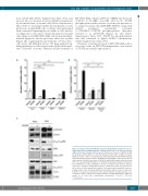
AXL and resistance to quizartinib in the hematopoietic niche
tions (10-20 nM) (Online Supplementary Figure S1C) and was not due to a decrease in active AC220 concentration by the metabolism of stromal cells (Online Supplementary Figure S1D). To investigate further the mechanism of such protection, stromal/AML cell co-cultures were performed with a transwell separating the two kinds of cells, thereby avoiding cell-to-cell contact. Transwell-separated stromal cells still protected FLT3-ITD AML cells from quizartinib- induced apoptosis, but the protective effect was weaker (Figure 1B), suggesting the involvement of both cell-to- cell contact and diffusible factors. We then analyzed sig- naling pathways in cells treated with AC220 in the pres- ence or absence of stroma. Whereas AC220 treatment of
AB
FLT3-ITD AML cell lines (MV4-11, MOLM-14) decreased Y591FLT3, T202/Y204ERK, Ser473AKT and Y694/Y699 STAT5 phosphorylation in the absence of stroma, the presence of co-cultured stromal cells (MS5, OP9, HS27A), along with AC220 treatment, specifically sustained Y694STAT5A/Y699STAT5B phosphorylation (hereafter referred to as pYSTAT5) (Figure 1C and Online Supplementary Figure S1E). STAT5 Tyr phosphorylation was still conserved at higher AC220 concentrations (Online Supplementary Figure S1F).
Therefore, stromal protection of FLT3-ITD AML cells is associated with an FLT3-ITD-independent conservation of STAT5 tyrosine phosphorylation.
C
Figure 1. Stromal cells protect MV4-11 cells from AC220-induced apoptosis and cor- relate with enhanced STAT5 activation. (A) MV4-11 FLT3-ITD acute myeloid leukemia (AML) cell line was incubated in the absence (Vehicle) or presence of AC220 (3 nM) for 48 hours, without (None) or with the indicated murine (OP9 or MS5) or human (HS27a) stromal cell lines. Cell apoptosis was determined by Annexin V/DAPI labeling followed by flow cytometry analysis. (B) MV4-11 FLT3-ITD AML cell line was incubated in the absence (Vehicle) or presence of AC220 (3 nM) for 48 h, without (None) or with OP9 cells separated (Stroma TW) or not (Stroma) by transwells. Apoptosis induction was determined as in (A). (C) MV4-11 cells were incubated in the absence (Vehicle) or presence of AC220 (1 nM), without (none) or with OP9 stromal cells (Stroma) for 48 h. Upon cell lysis, immunoblot analysis of the indicated protein with β actin as a loading control, each of protein-dedicated immunoblot without (none) or with (stro- ma) OP9 co-culture was performed on the same membrane. Results shown are rep- resentative of three experiments. Graphs show the mean±Standard Error of Mean of results of at least three independent experiments. *P<0.05; **P<0.01; ***P<0.001.
haematologica | 2019; 104(10)
2019
