
Page 83 - 2019_09-HaematologicaMondo-web
P. 83
A unique ABCB7-FECH-ABCB10 complex
(Figure 4A) and decreased Mfrn1 to levels comparable to those of cells treated with desferrioxamine (Figure 4A). A detectable compensatory activation of the IRE-binding activity of Irp1 in Irp2-KD cells (Figure 4B) was insuffi- cient to stabilize Tfrc to levels comparable to those in
controls. Mitochondrial iron levels decreased by more than 50% (Figure 4C), due to the decreased half-life of MFRN1 under the iron-limiting conditions generated by the lack of Irp2 (Figure 4D). Levels of radiolabeled hemo- globin dropped by more than 80% (Figure 4E,F), causing
F
A
B
C
D
G
E
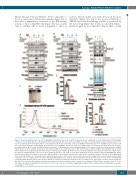
Figure 3. Heme biosynthesis defect in cells lacking Abcb7. (A) Protein blots to Irp2, Irp1, Tfrc, Hbb-b1, Hba-a2, and Bach1 in control (NT) and Abcb7-KD G1E-ER4 cells before (-β-estradiol) and after (+β-estradiol) 72 h of differentiation. The effect of the Alad inhibitor succinylacetone (SA) during cell differentiation was also test- ed. 55Fe incorporated into hemoglobin (Hb) in cells silenced for Abcb7 was profoundly decreased. (B) Mitochondrial fractions from G1E-ER4 cells treated as in (A) were probed with antibodies against Abcb7, Alas2, Abcb10, Fech, and Mfrn1. Tom20 was used as a loading control. (C) Cell pellets of control (NT β-estr) or Abcb7- knockdown (KD) (si-Abcb7 β-estr) G1E-ER4 cells differentiated for 72 h showed decreased hemoglobinization in cells depleted of Abcb7. (D) Representative oxidized UV-VIS spectra of heme, with the characteristic Soret band at 414 nm and additional peaks at 541 and 576 nm, in control and Abcb7-KD G1E-ER4 cells 72 h after differentiation showed significantly lower heme levels in cells depleted of Abcb7. (E) Heme levels in control and GIE-ER4 cells depleted of Abcb7 for 3 days. Values are means ± standard deviation. (F) Levels of heme-bound cytochromes c (Cyc) and c1 (Cyc1) were significantly decreased in Abcb7-depleted G1E-ER4 cells. Native immunoblots to Atp5a (CV subunit), Uqcrc2 (CIII subunit) and Mtco1 (CIV subunit) showed that levels of heme-containing complexes CIII and CIV were significantly decreased in Abcb7-KD cells. CIV activity, which requires two heme centers, was also decreased. Tom20 was used as a loading control. Peroxisomal fractions showed comparable levels of catalase protein in control and Abcb7-KD G1E-ER4 cells. Catalase (Cat) protein levels increased to the same extent in control and Abcb7-KD cells during differentiation. Pmp70 (also known as Abcd3) is a marker of peroxisomes and was used as a loading control. (G) Catalase activity in control and Abcb7- KD cells before and after differentiation showed profound defects in heme-dependent catalase activity in cells depleted of Abcb7. Values are expressed as % of con- trol. Values are means ± standard error of mean. (A-C, E-G, n=6; D, n=3). See also Online Supplementary Figure S15 for densitometries of immunoblots and statis- tical analyses. ***P<0.001
haematologica | 2019; 104(9)
1761
