
Page 84 - 2019_09-HaematologicaMondo-web
P. 84
N. Maio et al.
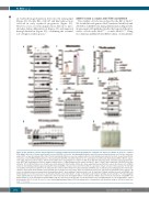
an evident hemoglobinization defect in cells lacking Irp2 (Figure 4G). Double KD of Abcb7 and Irp2 reduced iron overload in early erythroid progenitors (Figure 4C). However, it also severely impaired iron delivery to mito- chondria during differentiation (Figure 4C) and impaired hemoglobinization (Figure 4G), confirming the essential role of Irp2 in erythropoiesis.29
ABCB7 formed a complex with FECH and ABCB10
The stability of Fech was reduced by the KD of Abcb7. We found that endogenous Abcb7 interacted with Fech and Abcb10 in G1E-ER4 cells during differentiation (Figure 5A), in agreement with published studies that reported the inter- action of Fech with Abcb730-32 or with Abcb10.31,32 Using two-dimensional/Blue-Native(BN)/sodium dodecylsulfate
A
C
E
F
D
BG
Figure 4. Irp2 activation sustains mitochondrial iron overload mediated by mitoferrin-1 upregulation in erythroid cells depleted of Abcb7. (A) Irp2, Tfrc, ferritin H (Fth), Mfrn1, Alas2, Aco2, Hspa9 and Fam96b levels in G1E-ER4 cells before and during differentiation and either upon treatment with the iron chelator desferriox- amine (DFO) or upon knockdown (KD) of Irp2. Tom20 and tubulin (Tub) were used as loading controls for mitochondrial and cytosolic fractions, respectively. (B) Iron- responsive element (IRE)-binding activities of Irp1 and Irp2 in G1E-ER4 cells differentiated for 72 h and transfected with small interfering (si)-RNA to KD the expres- sion of Irp2, Abcb7 or both for 3 days. DFO-treated samples were run alongside to compare the KD effect with iron deficiency on the activation of iron regulatory pro- teins (IRP). Irp1 blotting did not show a change in protein levels. (C) Mitochondrial iron in control and Abcb7-, Irp2- or Abcb7/Irp2- double-KD G1E-ER4 cells before and after 72 h of differentiation. Loss of Irp2 reduced iron delivery to mitochondria. (D) A pulse-chase experiment was performed to assess the turnover rate of Mfrn1 under the iron-limiting conditions generated by KD of Irp2. G1E-ER4 cells were silenced for 72 h to KD the expression of Irp2. Cells were then pulsed for 30 min with 35S-Cys/Met, followed by incubation for the indicated time points in differentiation medium. Radiolabeled Mfrn1 was visualized by autoradiography after immunopre- cipitation and sodium dodecylsulfate polyacrylamide gel electropheresis (top panel), whereas total protein levels were assessed by immunoblot (lower panels). (E) Levels of radiolabeled iron incorporated into hemoglobin in Abcb7-, Irp2- and Abcb7/Irp2 double-KD cells. (F) Irp2, Abcb7, Mfrn1 and Hba-a2 levels in G1E-ER4 cells differentiated for 72 h in the presence of β-estradiol and silenced for either Abcb7, or Irp2 or simultaneously for Abcb7 and Irp2. A representative 55Fe-autoradiogram shows significantly decreased levels of radioactive iron incorporated into hemoglobin (Hb) in cells depleted of Abcb7, Irp2 or both. (G) Cell pellets of G1E-ER4 cells differentiated for 72 h and transfected with siRNA to KD the expression of Abcb7, Irp2 or both showed defective hemoglobinization in cells lacking Abcb7, Irp2 or both proteins. (A-D, F, G, n=4; E, n=6). See also Online Supplementary Figure S16 for densitometries of immunoblots and statistical analyses. NT: not treated (con- trol). ***P<0.001
1762
haematologica | 2019; 104(9)
