
Page 72 - 2019_09-HaematologicaMondo-web
P. 72
L. Crisafulli et al.
ABCD
E
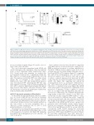
Figure 4. Inhibition of miR-127-3p function severely impairs hematopoietic stem cell (HSC) self-renewal. (A) Kaplan-Mayer survival curves of secondary recipients (n=10-11 from two independent experiments; Log-rank test) of 127DR- and empty vector (EV)-transduced cells. The arrow below the horizontal axis indicates the time of first peripheral blood (PB) withdrawal (4 weeks after transplant). (B-D) PB analysis four weeks after transplant (n=10-11 from two independent experiments). (B) FACS analysis for myeloid chimerism, measured as percentage of donor CD45.2+ cells within CD11b+ cells. (C) Percentage of myeloid and lymphoid cells within CD45+ donor gate determined by FACS analysis. (D) Platelet counts determined by hemocytometer analysis. (E) FACS analysis of 127DR stem and progenitor cells in one of the two secondary recipients that survived throughout the experiment. Dot plots are gated on LKS population and a representative sample is shown for the EV group. Histogram overlay (right) shows dGFP expression within the HSC (non-transplanted and EV) or the LKS (127DR) gate.
tion in secondary recipients (Figure 4C and D), in accor- dance with a stem cell defect.
Two out of eleven mice transplanted with 127DR cells survived throughout the experiment, with very different levels of donor chimerism (Online Supplementary Figure S3A). Several weeks after transplant, we analyzed the most primitive hematopoietic compartment within the BM of the two secondary sponge recipients that were still alive. This analysis revealed that HSC were either absent (Figure 4E) or belonged to the recipient (Online Supplementary Figure S3B). LKS multipotent progenitors were donor-derived GFP+, indicating that HSC had origi- nally engrafted at the time of the secondary transplant and were able to generate differentiated progeny throughout time; however, they failed to self-renew. Therefore, miR- 127-3p activity is crucial for the maintenance of phenotyp- ically and functionally defined HSC.
miR-127-3p prevents premature differentiation
To discern the biological mechanism at the basis of HSC loss, we tested if miR-127-3p is involved in regulating HSC quiescence, metabolic properties, survival, or preven- tion of premature differentiation, all necessary to preserve self-renewal. All these features were initially analyzed on Lin– cells isolated ex vivo from transplanted mice, as opposed to newly transduced cells, in order to avoid potential influences of cell culture.
The proportion of cycling and quiescent HSC was simi- lar in 127DR- and EV-transduced BM (Figure 5A), as well as the proportion of apoptotic cells (Figure 5B), indicating that miR127-3p downregulation does not alter HSC cell cycle or apoptosis.
Since oxidative stress has been described to negatively affect HSC function, we measured reactive oxygen species (ROS) production in response to cytokine stimulation, as described.23 ROS production induced upon short in vitro cytokine exposure was similar in HSC within Lin– cells iso- lated from 127DR or EV recipients (Figure 5C), suggesting that miR-127-3p downregulation does not lead to an increase in oxidative stress. However, after only one day of culture, a reduced expression level (measured by MFI) of the stem-cell associated cKit marker was observed in 127DR cells (Figure 5D), suggesting that miR-127-3p downregulation leads to accelerated differentiation. Lin– cells isolated from 127DR or EV recipients were also subjected to a standard colony assay (Figure 5E). 127DR stem and progenitor cells generated a significantly lower proportion of immature colonies compared to mature ones, in accordance with the hypothesis that they are more prone to differentiate. We confirmed this phenotype using freshly isolated HSC cultured in vitro. HSC were sorted from wt mice using the EPCR marker,24,25 trans- duced and analyzed at different time points. Single-cell colony assay performed early after transduction revealed that, despite the fact that the total number of colonies was comparable in the 127DR and EV groups, sponged cells generated fewer CFU-GEMM colonies, which originate from the most immature cells (Figure 5F). FACS analysis did not show any differences in the two groups when the conventional CD48, CD150 staining was performed (Online Supplementary Figure S4A); however, at day 6, the proportion of HSC expressing EPCR was reduced (Figure 5G and Online Supplementary Figure S4B). Moreover, a higher proportion of cells displayed early signs of differen-
1750
haematologica | 2019; 104(9)
