
Page 70 - 2019_09-HaematologicaMondo-web
P. 70
L. Crisafulli et al.
ABC
DE
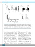
Figure 2. Expression of miR-127-3p. (A) Histogram shows fold difference (FD) of miR-127-3p expression in hematopoietic stem cell (HSC) from 3-4-week old Tie2Cre+Pbx1-/f mice relative to HSC from Tie2Cre-Pbx1+/f littermate controls, as measured by quantitative real-time polymerase chain reaction (qRT-PCR). Due to the extreme paucity of stem cells from the Tie2Cre+Pbx1-/f model,4 HSC from nine mice were grouped in two pools to perform qRT-PCR; bars indicate the range. (B) Expression of the indicated miRNA in HSC from 4-12-week old wild-type (wt) mice as measured by qRT-PCR and expressed in arbitrary units (AU). (C) Expression of the indicated miRNA in multi-potent progenitors (MPP) from wt mice relative to their expression level in HSC. (B and C) miR127-3p and let-7e, n=4; miR-99b, n=8; miR-125a and miR-125b, n=1; miR-221, n=6; miR-126a, n=2; n: number of pools (2-5 mice/pool). For miR-126, bars indicate the range. (D) miR-127-3p expression level in steady state bone marrow cell subpopulations from two pools of wt mice (2-3 mice/pool). (E) qRT-PCR analysis of miR-127 expression in human hematopoietic mature CD34– cells (or in CD34+CD38+ progenitors) relative to the CD34+ mobilized peripheral blood (MPB) or CD34+CD38- cord blood (CB) compartments. N=2 healthy donors for each source, bars indicate the range. All donors signed informed consent. When miRNA expression is indicated as FD, black bars indicate the sample relative to which FD is calculated.
transplanted mice (Online Supplementary Figure S2B). Similarly, the level of engraftment by sponge-transduced HSC (named 127DR for miR-127-3p functional downreg- ulation), measured as the proportion of cells expressing the donor marker CD45.2 over time, was comparable to that of HSC transduced with a control vector carrying only dGFP (named EV for empty vector) (Figure 3B) or with untransduced cells (data not shown), with the excep- tion of one mouse out of eleven. Sponge and control vec- tors express dGFP under the SFFV promoter, which is highly active in HSC, progenitors and myeloid cells, par- ticularly monocytes. Due to its rapid turnover, dGFP is only detectable when expressed at very high levels. Therefore, even though the SFFV promoter has good and modest activity in B and T lymphocytes, respectively, the dGFP marker is hardly detectable in these lineages.22 For this reason, we followed by FACS analysis dGFP expres- sion in monocytes; this also served as an indirect measure of dGFP expression in HSC since, due to their high turnover, myeloid cells are continuously generated from
HSC. Virtually all monocytes were GFP+, indicating a very efficient transduction of long-term reconstituting cells (Figure 3C). Importantly, GFP-negative or low B and T lymphocytes isolated from the spleen of transplanted mice contained vector sequences (Online Supplementary Figure S2C), indicating that they derived from transduced cells. We therefore safely monitored the presence of trans- duced cells in vivo through the donor marker CD45.2, since we could not rely on dGFP expression in all cells.
The kinetics of peripheral myeloid, B- and T-cell recon- stitution from donor cells was similar in mice transplanted with 127DR- or EV-transduced HSC, as well as the pro- portion and number of the different cell types (Figure 3D), suggesting that suppression of miR-127-3p activity did not affect multi-lineage reconstitution. However, FACS analy- sis of the BM at necropsy revealed a significant depletion of donor-derived phenotypically defined HSC, which were GFP+ (Figure 3E), without affecting their output of multi-potent progenitors and their lymphoid and myeloid differentiation potential, as confirmed on BM and spleen
1748
haematologica | 2019; 104(9)
