
Page 115 - 2019_09-HaematologicaMondo-web
P. 115
UBE2A somatic variants in CML progression
D
E
F
ABC
GH
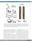
Figure 2. UBE2A silencing in K562 cells. (A) Real-time quantitative polymerase chain reaction (RT-qPCR) analysis of total RNA extracted from K562 cell lines infected with a lentiviral based system for UBE2A silencing (shNC: scrambled negative control; shUBE2A: UBE2A silenced cells). Values are normalized on shNC cells (***P<0.0001). (B) Western blot analysis of total cell lysates from K562_shNC and K562_shUBE2A cells. (C) Heat map of RNA-sequencing data showing color- coded expression levels of differentially expressed genes in three distinct populations of K562-shUBE2A compared to control (shNC). (D) RT-qPCR analysis in K562 cell lines of a subset of differentially expressed genes identified by RNA-sequencing. (E) Gene set enrichment analysis of the shUBE2A transcriptome. (F) RT-qPCR analysis in the 32Dcl3 cell line of a subset of differentially expressed genes identified by RNA-sequencing. (G and H) CSF3R protein levels in total cell lysate of K562 cells (G) and of BC/CP samples from patient #3, carrying UBE2A mutation in the BC phase (H).
samples at onset, 14 AP/BC, 40 acute myeloid leukemia, and 38 aCML samples. No evidence of UBE2A mutations could be found in the CP, AML or aCML samples, while in two AP/BC samples, somatic UBE2A variants D114Y and M34fs were detected. Globally, acquired UBE2A mutations could be detected in a total of 16.7% (4 of 24) advanced (AP/BC) CML cases (95%CI: 1.78-31.62) (Table 2).
UBE2A mutations affect protein activity
Polyphen-2 (http://genetics.bwh.harvard.edu/pph/),20
DANN11 and FATHMM-MKL21 analyses revealed that all the UBE2A variants identified were potentially damaging, as also suggested by the presence of a N-terminal frameshift variant (M34fs) in one of the patients (Table 2). To gain insight into the functional role of UBE2A muta- tions, we stably transfected the BA/F3_BCR-ABL1 cell line14 with the wild-type (WT) and the mutated UBE2A variants, I33M and D114V. The level of UBE2A expression
in stable transfectants was verified both at protein (Figure 1A) and mRNA (Figure 1B) levels.
The analysis of the levels of ubiquitin-conjugated H2A, a known UBE2A substrate,22 in total cell lysate revealed a decreased H2A ubiquitination for both UBE2A variants compared to WT (Figure 1C), with the effect of I33M being more prominent. In line with these findings, sug- gesting a decreased UBE2A activity for both variants, ubiquitination assay performed with in vitro translated WT and mutated UBE2A proteins confirmed a decrease in ubiquitin-conjugating activity for mutants compared to the WT form (Figure 1D). To further support this indica- tion, we developed a new in vitro assay based on the meas- urement of the AMP concentration as a proxy to assess the overall level of ubiquitination. This test was per- formed in the presence of GST-ubiquitin and of the E1 ubiquitin activating enzyme UBA1 together with WT or mutated UBE2A; this revealed a significant decrease in
haematologica | 2019; 104(9)
1793
