
Page 85 - 2019_08-Haematologica-web
P. 85
ZFP36L1 enhancer DNA methylation in MF
ed in enhancer regions of the genome. A preliminary analysis of the global DNA methylome revealed the absence of DNA methylation differences between pri- mary and secondary MF. This constitutes the first key finding of the present study and allowed us to use all the MF samples in a single cohort for further analysis. Primary and secondary MF are known to have very similar biolog- ical features, presenting symptoms and clinical course and in fact, both entities are treated indistinctively according to most published guidelines3, 31 Nevertheless, some recent evidence from large retrospective trials has suggested that traditional prognostic factors may not be applicable to sec- ondary MF as patients with post-ET MF seem to survive longer than those with post-PV MF or primary MF.3,31-33
A
The remarkably homogenous epigenetic profile of all our MF samples supports a common biological origin of pri- mary and secondary MF.34 The DNA methylomes of the novel MF subtypes defined by the new 2016 WHO classi- fication (prefibrotic and overt MF) remain to be character- ized and it will be interesting to establish whether these subtypes have different methylation profiles. This aspect exceeded the possibilities of our cohort (retrospective availability of histology samples) but warrants further investigation.
Although previous studies have already interrogated the DNA methylation landscape of MF,7 their findings are lim- ited to small numbers of epigenetic abnormalities mainly focused on the study of promoter regions. Our genome-
B
C
DEF
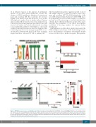
Figure 3. ZFP36L1 rescue decreases cell viability in myelofibrosis. (A) Consensus binding motif for ZFP36L1 obtained by DREME motif discovery among transcripts with putative AU-rich motifs upregulated in myelofibrosis samples. (B) Efficiency of infection measured by the percentage of EGFP-positive cells after lentiviral infec- tion. (C) Quantitative polymerase chain reaction validation of ZFP36L1 restoration in the SET-2 cell line after lentiviral infection. (D) ZFP36L1 protein restoration measured by western blot in the SET-2 cell line after lentiviral infection. (E,F) ZFP36L1 rescue with lentiviral vector infection in the SET-2 cell line decreased cell pro- liferation rate (E) and increased annexin V-positive cells (F).
haematologica | 2019; 104(8)
1577
