
Page 69 - 2019_07 resto del Mondo-web
P. 69
Mechanisms of NCOA4-maintained murine erythropoiesis
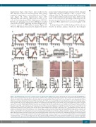
Supplementary Figure 2SC). Ncoa4rec mice reached lower RBC numbers, hematocrit and hemoglobin nadirs which required a longer time to recover than those of Ncoa4fl/fl mice (Figure 3A, Online Supplementary Figure S2D) although they did reach the same levels as those of con- trols at a later time-point (Online Supplementary Table S4). Reticulocyte percentage was lower in KO mice at initial time points after PHZ suggesting an initial sluggish reticu- locytosis response to PHZ-induced anemia (Figure 3B). However, KO animals demonstrated a higher reticulocyte
peak at day 10 and percentage reticulocyte level remained significantly elevated compared to that of controls at the endpoint (day 12). The trend observed in absolute reticu- locyte number (Online Supplementary Figure S2D) was the same as the reticulocyte percentage, indicating that the increase in reticulocytosis observed is not a consequence of decreased RBC number due to increased fragility of the cells.
A
BCDE
FGKLM
HIJ
Figure 3. Ncoa4-deficient mice show a less effective response to induced stress-erythropoiesis. Ncoa4rec and Ncoa4fl/fl mice were treated with phenylhydrazine to induce red blood cell lysis. Whole blood, tissue, and serum were analyzed. Panels C-M show measurements performed at the experimental endpoint (day 12), 8 days after the last administration of phenylhydrazine. (A) Serial complete blood count profiling of Ncoa4rec and Ncoa4fl/fl mice [5 mice/group, error bars represent the stan- dard error of mean (s.e.m)]. (B) Reticulocyte percentage from Ncoa4rec and Ncoa4fl/fl mice (5 mice/group, error bars represent the s.e.m). (C) Erythropoietin levels in serum of Ncoa4fl/fl (n=4) versus Ncoa4rec (n=5) mice (error bars show the s.e.m). (D) Increased Hif-2a protein levels in kidney from Ncoa4rec mice after PHZ adminis- tration. β-actin (Actb) served as a loading control. (E) Spleen size normalized to body weight is increased in Ncoa4rec mice at the endpoint (5 mice/group, error bars represent the s.e.m). (F) Ter119-Cd44 staining of bone marrow cells isolated from Ncoa4fl/fl and Ncoa4rec mice. Cells within the Ter119+ population were quantified and cells at each stage of differentiation are represented as the percentage of the total population (3 mice/group, error bars represent the s.e.m.) Stage I: proery- throblasts, stage II: basophilic and polychromatic erythroblasts, stage III: orthochromatic erythroblasts, stage IV: reticulocytes and stage V: mature red cells. (G) Representative field (20x) of 5 mice/group (scale bar=100 μm) of Cd68 staining in liver from Ncoa4fl/fl and Ncoa4rec mice. (H) Quantification (at least 5 fields per sample) of erythroblastic islands (5 mice/group, error bars represent the s.e.m.) associated with Cd68 staining. (I) Hamp mRNA fold-change in livers of Ncoa4rec and Ncoa4fl/fl mice (n=2/group, error bars represent the s.e.m.). (J) Erfe mRNA fold-change in bone marrow of Ncoa4rec and Ncoa4fl/fl mice (n=5 Ncoa4fl/fl, n=4 Ncoa4rec, error bars represent the s.e.m.). (K-M) Increase in tissue iron staining and levels in Ncoa4rec mice as determined by Prussian blue staining of spleen (K), liver (L) and kidney (M) (representative field of 5 mice/group, 10x, scale bar=200 μm). Bottom: Tissue iron levels in spleen, liver, and kidney (3 mice/group, error bars represent the s.e.m.). Statistical comparison was performed using a two-tailed Student t-test: *P<0.05, **P<0.01, ***P<0.001, except for panel B, where a one-tailed Student t-test was performed: *P<0.05. RBC: red blood cell count; HCT: hematocrit; HGB: hemoglobin; MCH: mean corpuscular hemoglobin; CHr: reticulocyte hemoglobin content; MCV: mean corpuscular volume; RDW: red blood cell distribution width; RETIC: reticulocyte count; PLT: platelet count; Epo: erythropoietin; b.w.: body weight.
Both Epo (Figure 3C) and Hif-2a (Figure 3D) levels were elevated in mutant animals, which also had enlarged
haematologica | 2019; 104(7)
1347
