
Page 59 - 2019_07 resto del Mondo-web
P. 59
NIPBL/NPMc+ interplay in myeloid differentiation
AB
C
DEF
GH
I
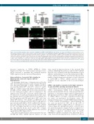
Figure 3. Canonical Wnt signaling is hyper activated in nipblb-MO and NPMc+ Tg(TOPdGFP) injected embryos at 3 days post-fertilization. (A, B) Quantitative reverse transcriptase polymerase chain reaction analyses of gfp and axin2 expression in nipblb-MO and NPMc+ Tg(TOPdGFP) injected embryos indicated an increase of canonical Wnt activation status in comparison to that in controls. (C) Scheme of the trunk-tail region of embryos. Confocal images were always taken of the same region of the embryo, comprising the tip of the yolk sack extension (YSE) between the dorsal aorta (DA, red line) and the vein (V, blue line) as indicated by pink brack- ets. (D-H) Confocal images of the caudal hematopoietic tissue (CHT) of Tg(TOPdGFP) embryos injected with nipblb-MO (E) and NPMc+ mRNA (G) showed an increase of GFP+ cells in comparison to controls (D). Co-injection of the Wnt inhibitor dkk1b mRNA rescued the number of GFP+ cells (F-H). (I) Quantification of GFP+ cells in the selected region of the CHT. Images were processed as described in the Online Supplementary Methods. The scale bar represents 100 μm. *P<0.05, **P<0.01 and ***P<0.001. ns: non-significant; MO: morpholino; GFP: green fluorescent protein; ctrl: control.
increased expression of NIPBL (NIPBL>1) (Online Supplementary Figure S7). Further analyses in larger cohorts will be necessary to determine the correlation between NIPBL expression and the canonical Wnt pathway.
Hyper-activation of canonical Wnt signaling in hematopoietic stem cells impairs myeloid differentiation
To determine which type of hematopoietic cell in the CHT showed hyper-activation of the canonical Wnt path- way following nipblb-MO or NPMc+ mRNA injections, we sorted the CD41:GFPlow cells (0.8% control, 1% nipblb- MO, 1.1% NPMc+) from 3 dpf embryos injected with nip- blb-MO or NPMc+ mRNA and observed a modest increase in axin2 expression in these cells in comparison to the expression in CD41:GFPlow cells from controls (Figure 4A- A’’’). Moreover, we performed immunofluorescence stain- ing with the Active β-catenin (Active β-cat) antibody for the Wnt pathway and GFP antibody for HSC cells of the CD41:GFP embryos. The number of GFP/Active β-cat double-positive cells present in the CHT of 3-dpf embryos injected with nipblb-MO or NPMc+ mRNA (5 double-pos- itive cells indicated by the arrows in the CHT of Figure 4C’’-D’’) was higher than that in controls (2 double-posi- tive cells indicated by the arrows in the CHT of Figure 4B’’), demonstrating that the Wnt pathway was activated specifically in HSC (Figure 4B-D’’).
The myeloid differentiation defects presented by nip- blb-MO and NPMc+ mRNA injected embryos at 3 dpf
were caused by hyper-activation of the canonical Wnt pathway. Inhibiting this pathway, either by dkk1b mRNA injection or by treatment with the Wnt pharmacological inhibitor indomethacin,26 rescued the hematopoietic phe- notype. Indeed, by WISH we showed that the increased number of myeloid precursors positive for spi1b observed in nipblb-MO or NPMc+ mRNA injected embryos, returned to levels comparable to those in controls (Figure 5A-G).
NPMc+ and nipblb cooperation in the hyper-activation of canonical Wnt signaling and myeloid defects
The evidence that we had collected so far showed that NPMc+ downregulates NIPBL, both in human AML patients and in zebrafish. We also demonstrated that dys- regulation of both genes impairs myeloid differentiation through hyper-activation of the canonical Wnt pathway. To further address a possible cooperation between the two genes, we performed dose-response assays. We injected subcritical doses of nipblb-MO (0.6 pmol/embryo) or NPMc+ mRNA (50 pg/embryo), which singularly did not cause an increase in GFP+ cells in the CHT of Tg(TOPdGFP) embryos (Figure 6B,C) in comparison to controls (Figure 6A, quantification in E), or in spi1b-posi- tive myeloid progenitors (Figure 6G,H) in comparison to the number of cells in controls (Figure 6F, quantification in J). When co-injected, subcritical doses of nipblb- MO/NPMc+ mRNA recapitulated the phenotype previ- ously observed following injections of full doses, with an
haematologica | 2019; 104(7)
1337
