
Page 142 - 2019_07 resto del Mondo-web
P. 142
A. Uchida et al.
(Figure 4B). Although the expression levels of MCL1 were nearly equal between BJAB and Karpas231 cells, S63845 failed to exert additive pro-apoptotic effects in Karpas231 cells. These results indicate that the intrinsic anti-apoptot- ic activities in BJAB are equally dependent on BCL2 and MCL1, but those in the two DH-DPL-cell lines and SU- DHL10 cells depend mainly on BCL2 and MCL1, respec- tively. We, therefore, focused on the biological effects of venetoclax especially in the two DH-DPL cell lines.
Venetoclax leads to dephosphorylation of BCL2 and downregulates MCL1 expression in DH-DPL cells
We then examined alterations of protein levels after exposure to venetoclax. Western blot analysis showed that expression levels of BCL2 were unchanged at least 6 h after exposure to venetoclax in Karpas231 and OCI-Ly8 cells (Figure 5A,B). In contrast, phosphorylation of BCL2 was clearly downregulated, and expression levels of BIM were also decreased 3 h after exposure to venetoclax in both DH-
A
B
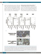
Figure 1. Expression of MYC and BCL2 family proteins in germinal center B-cell-like diffuse large B-cell lymphoma. (A) Immunohistochemistry-detected protein expression of MYC, BCL2, MCL1, BIM and BAX in clinical samples of double-hit (DH) high grade B-cell lymphoma with MYC and BCL2 rearrangements (HGBL) and other germinal center B-cell (GCB)-like diffuse large B-cell lymphomas (DLBCL) including non-DH double-protein-expression lymphoma (DPL). Straight bars represent the mean of each positive rate. The differences in positivity were calculated using the Mann-Whitney U test* and Student t test**. The positive rates of MYC and BCL2 were significantly higher in DH-HGBL than in other GCB-like DLBCLs (P=0.028 and P=0.013, respectively). In contrast, MCL1 was significantly less expressed in DHL-HGBL than in other GCB-like DLBCL (P=0.005). This significance was also observed between DH-HGBL and non-DH DPL (P=0.019). P values are described only when each statistical power was above 0.8. (B) MCL1 was stained mainly in the cytoplasm in all eight cases with DH-HGBL, whereas eight of 19 (42%) other GCB-like DLBCL cases, including non-DH DPL, showed the nuclear and cytoplasmic staining pattern.
1420
haematologica | 2019; 104(7)
