
Page 194 - 2019_04-Haematologica-web
P. 194
P. Durigutto et al.
other than β2GPI are expressed on the endothelial cell sur- face and represent potential targets for human IgG, we set out to determine whether the fluorescence was due to the IgG specifically directed against β2GPI. To do this, we set up a β2GPI-dependent CL assay in which the β2GPI sup- plementation was carried out by adding human purified β2GPI at increasing concentrations instead of fetal calf serum. The system allowed us to test the IgG reactivity with β2GPI added at different concentrations to the CL- plates. The anti-D1 IgG reacted with the β2GPI molecule most likely by recognizing the D1 epitope exposed on the β2GPI molecule following its binding to CL (Figure 4). The IgG level detected in the assay varied in different patients and was related to the concentration of β2GPI used to coat CL. In contrast, anti-D5 IgG failed to interact with CL- bound β2GPI even at the highest concentration of β2GPI, suggesting that D5 domains were not accessible to the antibodies under these experimental conditions. Like the anti-D5 antibodies, in the assay, the IgG from control sera were negative.
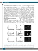
Figure 2. Anti-domain (D) 5 antibodies fail to induce thrombi in rats. Thrombus formation and vascular occlusion visualized by intravital microscopy in the ileal mesentery of rats that received an intraperitoneal injection of lipopolysaccharide (LPS) (2.5 mg/kg body weight) followed by the injection into the carotid artery of antibodies (10 mg/rat) directed against domain 5 (D5), domain 1 (D1), or anti-β2glycoprotein I (β2GPI)-negative (NHS). The number of thrombi (A) and vessel occlu- sions (B) were evaluated at various time intervals on 3 rats per each serum. The results are expressed as a ratio between the number of thrombi and the number of microvessels examined and as a percentage of occluded microvessels. Data are reported as mean±Standard Deviation. (C) Sections of the ileal mesentery show- ing endovascular thrombi in anti-D1-treated rat and undetectable in the vessels of animals receiving anti-D4/5-positive or anti-β2GPI-negative sera. Original magni- fication 100x. Scale bar 50 mm.
Antibodies to domain 5 interact with soluble β glycoprotein I
tic epitopes on D5, though this has not been formally proven.35 We first decided to examine the in vivo interac- tion of the antibodies with circulating β2GPI and the effect of this interaction on β2GPI bound to vascular endotheli- um. To this purpose, the in vivo model was slightly modi- fied administering IgG intraperitoneally followed 15 h later by LPS given by the same route; this approach would allow sufficient time for the antibodies to react with the target antigen prior to the binding of β2GPI to vascular endothelium promoted by LPS. The IgG from two sera with relatively high levels of antibodies to D1 and D5, respectively, and from an anti-β2GPI-negative serum were tested and the amount of vascular deposits of β2GPI and IgG was evaluated. As expected, the rat treated with anti- D1 developed endovascular thrombi associated with dep- osition of IgG, both of which were undetectable in ani- mals that received anti-D5-positive or anti-β2GPI-negative IgG (Figure 5). Analysis of the ileal mesentery showed that β2GPI was present on the vascular endothelium of the ani- mals that received the three IgG fractions with no clear difference in the staining intensity observed in the rats treated with anti-D5 and anti-D1 IgG (Figure 5).
Since the in vivo data did not provide convincing evi- dence of the ability of anti-D5 to prevent binding of circu- lating β2GPI to vascular endothelium, we decided to fur- ther investigate this issue using an in vitro inhibition assay. IgG purified from anti-D5-positive, anti-D1-positive or anti-β2GPI-negative sera were incubated with increasing
2Electron microscopy studies have revealed that β2GPI adopts a circular form in plasma and that this is main- tained by the interaction of D1 with D5.34 This special conformation prevents the access of autoantibodies to hidden epitopes on D119 and predicts the presence of cryp-
ABC
822
haematologica | 2019; 104(4)
