
Page 193 - 2019_04-Haematologica-web
P. 193
Anti-β2GPI-D5 are not thrombogenic in animals
AB
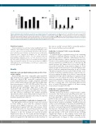
Figure 1. Anti-domain (D) 4/5 antibodies specifically react against domain (D) 5 of β2glycoprotein I (β2GPI). Reactivity of 5 anti-D4/5-positive patient sera (P1-P5) against different recombinant human β2GPI domains. (A) Reactivity against the combined D4/5 peptides ( ), in an assay produced for research use (QUANTA Lite β2GPI D4/5 ELISA, INOVA Diagnostics). (B) Reactivity against the recombinant domain D4 ( ) or D5 ( ) antigens separately immobilized on the wells of γ-irradiated polystyrene plates in in-house ELISA. Optical Density (O.D.) values are expressed as mean±Standard Deviation. Data were analyzed with the Student t-test for paired data. The average reactivity against D5 is significantly higher than that against D4 (P=0.0428).
Statistical analysis
Statistical analysis was performed using GraphPad Prism 6.0 for Windows. The domain reactivity of the anti-β2GPI D4/5 positive sera was expressed as mean+Standard Deviation (SD) and ana- lyzed with the paired Student t-test. Data from in vivo thrombus formation were compared by Dunnett test. The interaction between IgG and β2GPI bound to CL was analyzed with the Kruskall-Wallis with Dunn post-hoc test. The interaction between IgG and soluble β2GPI was expressed as median and interquartile range and analyzed with the two-way repeated measure ANOVA with Sidak post-hoc test. Probabilities of <0.05 were considered sta- tistically significant.
Results
the various sera IgG towards D4/5 is essentially similar to that observed in their reaction with D5.
Antibodies to domain 5 fail to cause thrombus formation in vivo
To evaluate the pro-coagulant activity of sera containing antibodies to different domains of β2GPI, two groups of serum IgG positive for either D1 or D5 domains were ana- lyzed for their ability to induce thrombus formation fol- lowed in vivo by intravital microscopy. IgG from sera neg- ative for antibodies to β2GPI served as a control group. All anti-D1-positive IgG induced blood clots that could be seen from 15 min after serum infusion (Figure 2). Their number progressively increased to reach the highest value after 1 h and was maintained thereafter for up to 90 min. Thrombus formation was associated with vascular occlu- sion that resulted in a marked decrease, and, in some ves- sels, in a complete blockage of blood flow. Conversely, the anti-D5-positive IgG did not exhibit pro-coagulant activity and failed to cause reduced blood flow. The latter results were not statistically different from those of anti-β2GPI- negative blood donors at each time point. On the contrary, the data of anti-D1 IgG were statistically different from those of anti-β2GPI-negative samples at all times starting from 15 min of analysis (P<0.05).
Antibody to phospholipid-binding protein profile of the serum samples
Anti-β2GPI IgG titers were comparable in the anti-D4/5- and anti-D1-positive samples [1.04±0.26 Optical Density (OD) and 1.46±0.48 OD, mean+SD, respectively]. The isolated anti-D4/5-positive samples displayed anti-D4/5 levels of 50.67±9.86 arbitrary units (AU) (mean±SD) while they were negative for aCL (<10 GPL) and LA. The isolat- ed anti-D1-positive samples showed anti-D1 levels of 75.36±17.15 AU (mean±SD), high titers of IgG aCL (124.4±46.9 GPL, mean±SD), and displayed LA activity. Control samples were negative in all the assays. The puri- fied IgG fractions maintained the antigen specificity shown in the whole serum. Clinical and serological data of all the subjects/patients included in the study are reported in Online Supplementary Table S1.
surface-bound β glycoprotein I 2
Fine epitope-specificity of antibodies to domains 4/5
The IgG against D4/5 used in this study were selected for their ability to react with the combined domains obtained from INOVA Diagnostics, but it was unclear whether they recognized one or the other domain or both. To clarify this point, we assessed the reactivity of serum IgG towards recombinant D4 and D5. The amino acid sequences of the two domains are reported in Online Supplementary Figure S1. The results presented in Figure 1 clearly show that all the anti-D4/5 reacted with D5 and did not recognize D4. The difference in the reactivity of
Having observed an absence of intravascular coagula- tion in rats that had received anti-D5-positive IgG, we decided to investigate whether this was due to the inabil- ity of the antibodies to interact with endothelium-bound β2GPI. To this end, samples of ileal mesentery were ana- lyzed for the presence of β2GPI, human IgG and C3. As expected from our previous findings,30 β2GPI was detected on the vessel endothelium of rats primed with LPS (Figure 3), while it was totally absent in unprimed animals (data not shown). A search for IgG and C3 revealed marked gran- ular deposits of both proteins on endothelial cells of rats treated with anti-D1 IgG, while a milder linear staining for IgG and absence of C3 were observed in rats receiving anti-D5 IgG (Figure 3). The animals treated with anti- β2GPI-negative sera showed negligible staining for IgG and undetectable C3 (Figure 3). Since several molecules
Antibodies to domain 5 fail to interact with
haematologica | 2019; 104(4)
821
