
Page 142 - 2019_04-Haematologica-web
P. 142
R. Burkhard et al.
way. Dysregulation of the NF-kB pathway is the onco- genic hallmark of ABC-DLBCL and PMBL.14 Of note, in 420 primary DLBCLs, high TIRAP expression correlated with poor survival and was significantly increased in high risk patients (data generated by SurvExpress32) (Online Supplementary Figure S2A). Besides, amplifications of 11q and 11q24 which also contain TIRAP, have been reported in approximately 20% of DLBCLs and PMBLs.11,33,34 Somatic TIRAP mutations occurring in various cancers including DLBCL are listed in COSMIC and genomic sequencing studies on several hundred DLBCLs revealed
somatic alterations in TIRAP in roughly 0.5% of cases.34,35 However, the role of TIRAP in tumorigenesis has so far not been investigated. Hence, we functionally investigate whether the identified TIRAP variant in this family con- tributes to lymphomagenesis.
TIRAP p.R81C variant is a potential novel risk factor for lymphomas
Whole exome sequencing revealed a heterozygous vari- ant within the coding exon 5 of TIRAP (c.241C>T) in both sisters and their Japanese mother. The variant was absent
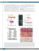
A
BC
DE
Figure 3. The JAK-STAT pathway has somatic mutations in multiple genes and is constitutively active. (A) aCGH probe view of the 9p24 gain. A duplication of the JAK2 locus was detected in both samples, shown as an increase of the average log2 ratio above zero (bold line). Shaded area indicated the extent of a copy number aberration. (B) Fluorescence in situ hybridization (FISH) signals with BAC probes for the 5’ and 3’ regions of JAK2. Arrows indicate examples of cells with multiple FISH signals. Note green autofluorescent sclerosing bundles in the background. Scale bars: 10 mm. (C) Array comparative genomic hybridization (aCGH) probe view of gains in 12p13 region, which among other genes also affect STAT2 and STAT6 as indicated by arrows. (D) (Top) Schematic representation of the human STAT6 gene locus with open and closed boxes indicating non-coding and coding exons, respectively. (Bottom) Confirmed somatic missense mutations located within the DNA binding domain of STAT6. Protein domain annotation according to Pfam. (E) The expression of phosphorylated (p) pJAK2, pSTAT3 and pSTAT6 in the two lym- phomas was assessed by immunohistochemistry. Scale bars: 10 mm.
770
haematologica | 2019; 104(4)
