
Page 140 - 2019_04-Haematologica-web
P. 140
R. Burkhard et al.
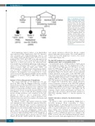
Figure 1. Mediastinal B-cell lym- phomas arising in 2 female siblings. Pedigree of the Swiss/Japanese fam- ily under study. Circles and squares represent female and male family members, respectively. Open symbols indicate unaffected persons; closed symbols repre-sent the 2 siblings affected by primary mediastinal B-cell lymphoma (PBML) and mediastinal non-germinal center (GC) diffuse large B-cell lymphoma (DLBCL), not otherwise specified (NOS), respec- tively. The deceased individual (sister 1) is marked by a slash through the symbol. The year of diagnosis is shown in brackets and the year of birth is denoted by *. Both lym- phomas were classified according to the World Health Organization 2017 classification.1
Both lymphomas lacked evidence of an Epstein-Barr virus infection (Online Supplementary Table S2), showed a clear cytoplasm and compartmentalizing sclerosis, and were CD20, CD30 and Ki-67 positive. However, the GC markers BCL6, CD10 and GCET1 were only expressed in the PMBL of sister 1, as was CD23 and BCL2. Despite the expression of CD30 and sclerosis, the clinical presenta- tion, morphology and immunophenotype of the tumor in sister 2 were consistent with a non-GC DLBCL NOS.1 Given its genetic (9p24 and 12q13 gains, SOCS1 and STAT6 mutations), and phenotypic characteristics (expres- sion of CD30, overexpression of JAK2-STAT-cascade members), this lymphoma can retrospectively be consid- ered to most probably represent a PMBL, and was initially designated as non-GC DLBCL NOS with features of PMBL.
Analysis of the coding genome of lymphomas
The somatic landscape of both lymphomas was ana- lyzed by WES using the Illumina technology on DNA derived from laser-dissected FFPE tissue sections (Online Supplementary Methods). Mutations were validated using Ion Proton sequencing, and their somatic origin was con- firmed by the absence in matched normal DNA isolated from PBMCs. A total of 192 and 130 confirmed clonal protein altering mutations were identified in the lym- phomas of sisters 1 and 2, respectively (Online Supplementary Table S7). Most of those mutations were missense mutations, with a low number of nonsense and splice site variants (Figure 2A).
In addition, somatic copy number alterations (CNA) were analyzed by array comparative genomic hybridiza- tion (aCGH). While the tumor of sister 1 contained seven gains and two deletions, three gains were detected in the lymphoma of sister 2. Interestingly, 9p24 and 12q13 gains were present in both lymphomas (Figure 2B). Fluorescence in situ hybridization (FISH) analyses revealed a trisomy at 8q24 (including MYC) in the lymphoma of sister 1, in line with the gain on chromosome 8 by aCGH (Figures 2B and 4B). Taken together, the analysis of CNA
and somatic mutations reflected the known complex genetic landscape in those entities. The overall number of somatic lesions in the lymphoma of sister 1 was consid- erably higher (Figure 2C).
The JAK-STAT pathway has somatic mutations in multiple genes and is constitutively active
As mentioned above, aCGH revealed a 9p24 gain in
both lymphomas (Figure 3A). The amplification of JAK2,
a key target of the 9p24 gain, in both tumors was con-
firmed by FISH (Figure 3B). Moreover, a gain of 12q13
encompassing STAT2 and STAT6, was detected in both
lymphomas (Figure 3C). Besides CNA, we also identified
somatic mutations in key genes of the JAK-STAT path-
way. Each tumor harbored a private missense mutation
within the DNA binding domain of STAT6 (Figure 3D). In
addition, SOCS1, encoding a negative regulator of the
JAK-STAT pathway, was mutated in the lymphoma of sis-
ter 1 (Figure 4A). Collectively, these somatic alterations
caused constitutive activation of the JAK-STAT pathway
as evidenced by high cytoplasmic expression of phospho-
rylated JAK2 (pJAK2) and increased nuclear pSTAT3 and
pSTAT6 expression in most tumor cells (Figure 3E). In
summary, despite distinct pathological and clinical fea-
tures, these data revealed a shared aberrant activation of
JAK-STAT signaling which is a known signature in PMBL.10,22,23
Genetic alterations related to the distinct clinical outcome
In contrast to ABC- and GCB-DLBCL, PMBLs have a favorable prognosis when responding to chemo- immunotherapy.2 To investigate genetic lesions associated with the different clinical outcome, we focused on genes with a reported pathogenic role in lymphomas and/or genes which were mutated in more than 10% of DLBCLs.4-10 Mutations in B2M, and TP53, REL and MYC gains as well as a CIITA break apart were confined to the PMBL of sister 1 who died of primary progressive disease (Figure 4A and B). Although amplification of PDL1 result-
768
haematologica | 2019; 104(4)
