
Page 117 - 2019_04-Haematologica-web
P. 117
Autophagy drives ETV6-RUNX1-positive leukemia
expression was further enhanced by co-expression of CBFβ (13-fold compared to control, P≤0.05). These results demonstrate that, although both RUNX1 and ETV6- RUNX1 function as transcriptional activators of Vps34, ETV6-RUNX1 induces Vps34 promoter activity more effi- ciently (Figure 2C). In addition, these results demonstrate that CBFβ acts as a co-activator for ETV6-RUNX1 in inducing Vps34 promoter activity. Additional luciferase reporter assays revealed that the ETV6-RUNX1 target genes HEY1, EGR1 and GATA1 induce Vps34 promoter activity in a dose-dependent manner. HEY1, EGR1, and GATA1 induced Vps34 promoter activity up to 13-fold (P≤0.05), 5.0-fold (P≤0.001), and 10-fold (P≤0.05), respec- tively (Figure 2C). GATA2 expression did not induce luciferase expression, which was in concordance with the absence of a GATA2 DNA binding domain in the -1338/+58 promoter region used for the reported assays (Figure 2A and C). Together, these results demonstrate that the Vps34 promoter is not only positively regulated by the ETV6-RUNX1 fusion protein itself, but also by its target genes HEY1, EGR1 and GATA1.
Autophagy levels are high in ETV6-RUNX1-positive BCP-ALL and regulated by ETV6-RUNX1 and Vps34
Since Vps34 is a key player in autophagy regulation, we hypothesized that ETV6-RUNX1-mediated upregulation of Vps34 induces autophagy in BCP-ALL cells. To investi-
gate this, autophagy levels were determined in ALL cell lines and primary BCP-ALL samples by western blot analysis and RPPA. Western blot analysis in a panel of ALL cell lines revealed that Vps34 protein levels are the highest in the ETV6-RUNX1-positive cell line REH (Figure 3A). In comparison to ETV6-RUNX1-negative ALL cell lines, lower levels of p62 (SQSTM1) and LC3B, both specifically degraded by autophagy, were observed in REH cells. These results suggest high levels of autophagy in REH cells (Figure 3A). RPPA on samples from a large cohort of newly diagnosed BCP-ALL patients revealed that the level of Vps34 was 9.6-fold higher in BCP-ALL cells compared to healthy bone marrow-derived mononuclear cells (59 BCP-ALL patients vs. 10 healthy controls; P≤0.001) (Online Supplementary Figure S6C). Vps34 levels were also signifi- cantly higher in ETV6-RUNX1-positive patient cells in comparison to ETV6-RUNX1-negative BCP ALL (B-Other) patient cells (1.2-fold, P≤0.05) (Figure 3B). In line with this, lower p62 protein levels were observed in ETV6-RUNX1- positive in comparison to ETV6-RUNX1-negative BCP- ALL patient cells (1.4-fold, P≤0.01) (Figure 3B). These RPPA data were confirmed by western blot analysis in a smaller set of patients (n=11 patients) (Online Supplementary Figure S6A and B).
Next, an siRNA approach was used to investigate whether ETV6-RUNX1 regulates autophagy. To quantify the level of autophagy, the number and volume of LC3B-
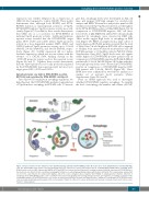
Figure 7. Proposed model for the induction of the Vps34-autophagy pathway in ETV6 RUNX1-positive B-cell precursor acute lymphoblastic leukemia (BCP-ALL) cells. The ETV6-RUNX1 fusion protein can transcriptionally induce the expression of various transcription factors, including GATA1, GATA2, HEY1, and EGR1. ETV6- RUNX1 and its co-factor CBFβ, together with GATA1, HEY1 and EGR1 can activate the Vps34 promoter, resulting in enhanced Vps34 expression in ETV6-RUNX1-pos- itive leukemic cells. Vps34, in turn, can initiate autophagy by forming a core autophagy-regulating complex with Beclin 1 and Vps15. This complex plays an important role in: 1) the early initiation (in complex with Atg14L); and 2) the vesicle elongation phase (together with UVRAG) of autophagosome formation. Induction of autophagy allows ETV6-RUNX1-positive cells to maintain homeostasis by degrading and recycling damaged proteins and organelles. In addition, activation of the autophagy program in ETV6-RUNX1-positive cells results in enhanced proliferation, survival and drug resistance. Inhibition of autophagy in ETV6-RUNX1-positive cells, by treatment with hydroxychloroquine (HCQ) or knockdown of Vps34, is sufficient to reduce proliferation and survival of leukemic blasts and to induce sensi- tization to L-Asparaginase.
haematologica | 2019; 104(4)
745
