
Page 135 - 2019_02-Haematologica-web
P. 135
Immunoprofiling and survival in PTL
phamide, doxorubicin, vincristine, and prednisone (R- CHOP) immunochemotherapy, CNS prophylaxis, and locoregional radiotherapy or surgery of the remaining con- tralateral testicle. While the addition of rituximab to sys- temic chemotherapy has improved the overall prognosis of DLBCL, treatment responses in PTL have not been equally evident.1,2
The tumor microenvironment (TME) and limited immune surveillance play important roles in the lym- phoma pathogenesis and survival.3,4 The TME of B-cell lymphomas consists of immune cells [e.g. T cells, macrophages, natural killer (NK) cells], stromal cells, blood vessels, and extracellular matrix. The TME may confer effective tumor recognition and clearance, but on the other hand, can also provide a protective niche for the tumor cells, and facilitate cell proliferation and survival.5 The interactions between lymphoma cells and the TME contribute to the ability of tumor cells to escape host immune surveillance; these mechanisms include loss of expression of human leukocyte antigen (HLA) class I and II molecules that interferes with correct tumor cell recog- nition by the cellular immune system and may provide tumor cells with an escape mechanism.6 Several studies have reported loss of HLA I and II expression in B-cell lymphomas, including PTL.7-11 Another immune escape mechanism is the expression of immune checkpoint mol- ecules, such as programmed cell death 1 (PD-1) and the cytotoxic T-lymphocyte-associated antigen 4 (CTLA-4).6
In this study, we aimed to characterize cellular and molecular immunological profiles in PTL, and associate the findings with outcome. We identified a gene signature enriched for T-cell- and NK-cell-related genes, the low expression of which predicts high risk of recurrence and mortality in patients with PTL and DLBCL. Our results emphasize the key role of the TME and immune escape in regulating therapy resistance in aggressive B-cell lym- phomas.
Methods
Patients' characteristics and samples
The study consisted of formalin-fixed paraffin-embedded (FFPE) samples from primary orchiectomy of 60 patients diag- nosed with PTL in 1993-2013 (Table 1). Twenty-eight of the patients were diagnosed and treated in the pre-rituximab era with anthracyclin-based chemotherapy, whereas 32 of the patients were treated with rituximab-containing immunochemotherapy. In addition, 34 patients received CNS prophylaxis, consisting mainly of intravenously administered high-dose methotrexate and/or high-dose cytarabine. Nineteen patients received treatment of the contralateral testicle (radiotherapy or orchiectomy). The cell-of-origin (COO) was determined by immunohistochemistry using the Hans algorithm.12 The study was approved by the Ethics Committees in Helsinki and Tampere, Finland, and by the Finnish National Authority for Medicolegal Affairs, and by the Institutional Review Boards of the institutes involved in the study.
The dataset of primary DLBCL from the Cancer Genome Characterization Initiative (CGCI) (the database of Genotypes and Phenotypes study accession: phs000532.v2.p1; n=96)13 was used for comparison (Table 1).
Gene expression analysis
Gene expression from the FFPE samples of 60 PTL patients was profiled with the Nanostring nCounter Human PanCancer
Immunoprofiling Panel (XT-CSO-HIP1-12, NanoString Technologies, Seattle, WA, USA). The detailed protocol is provid- ed in the Online Supplementary Methods.
Immunohistochemistry
Formalin-fixed paraffin-embedded tissue microarray (TMA) was utilized for the immunohistochemical (IHC) analyses. The protocol for HLA-ABC, HLA-DR, and β2 microglobulin staining is provided in the Online Supplementary Methods. Membranous staining in the majority (>90%) of tumor cells was scored as nor- mal (highly positive). Cases with mixed cytoplasmic and mem- branous staining were scored as moderately positive. Cases with
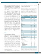
Table 1. Patients’ characteristics of the primary testicular lymphoma (PTL) and diffuse large B-cell lymphomas (DLBCL).
Characteristics
Number of patients
Age
Men
Women
Molecular subgroup
GCBa non-GCB nd
Stage I-IIb
III-IV
IPI score
0-2
3-5
Elevated LDH CNS prophylaxis
IV-prophylaxis
IT-prophylaxis
Treated with rituximab-containing regimen Primary CNS involvement
Relapses
Primary refractoryc
CNS relapse/progression Systemic and CNS relapse Systemic relapse/progression
Late relapse CNS relapse
Systemic and CNS relapse
Systemic relapse
Deaths
Lymphoma-specific
Other
PTL cohort All n (%)
60 (100)
16 (27)
44 (73) 60 (100)
15 (25) 40 (67) 5 (8)
39 (65) 21 (35)
42 (70)
18 (30) 22 (37) 34 (57) 31 (52) 8 (13) 32 (53) 2 (3) 18 (30) 11 (18) 2 (3)
2 (3) 7 (12) 7 (12) 2 (3) 0 (0) 5 (8) 25 (42) 16 (27)
9 (15)
DLBCL cohort All n (%)
96 (100)
41 (43)
55 (57)
63 (66) 33 (34)
55 (57) 41 (43)
18 (19) 78 (81)
25 (26)
71 (74) n/a n/a n/a n/a 91 (96) n/a 27 (28) n/a n/a n/a n/a n/a n/a n/a n/a 24 (25)
21 (22)
3 (3)
<60 years
≥60 years Sex
haematologica | 2019; 104(2)
aGCB; germinal-center B cell; nd: not determined; LDH: lactate dehydrogenase; n/a: not assigned; CNS: central nervous system; IV: intravenous; IT: intrathecal. bIsolated bilateral testicular involvement was defined as stage IAE.cRelapse/progression less than 12 months after the diagnosis and/or within 6 months from the end of primary treatment.
339
