
Page 221 - 2019_01-Haematologica-web
P. 221
The impact of plasma on cold storage of APC
pendent manner. While similar Annexin V binding was observed on PLTs from PAS-35-APCs and Plasma-APCs stored at both temperatures, cells stored in 20% residual plasma (PAS-20-APC) showed higher exposure of the apoptosis marker phosphatidylserine, particularly when PLTs were cold-stored (Figure 2D).
Effect of residual plasma on adhesion of cold-stored platelets
The adhesion of TRAP-activated PLTs was analyzed after seven days of storage. More PLTs from APCs stored at 4°C adhered to collagen and fibrinogen compared to PLTs stored at RT (Figure 3A). This effect was statistically significant for PLTs stored in 100% and 35% plasma (Figure 3B and D). Moreover, the individual percentage of each spreading pattern (type 1, 2, 3 and 4) (Online Supplementary Figure S2) was always higher after cold stor- age, regardless of plasma volume, but without reaching statistical significance (Online Supplementary Table S1A and B). Cells stored in the presence of 35% plasma showed similar adhesive response to both proteins compared to 100% plasma (Figure 3B and D). In contrast, significantly fewer PLTs adhered to both proteins from cold-stored PAS-20-APCs compared to plasma-APCs (percentage of adherent PLTs to collagen, mean±SEM: 13±4 vs. 41±4, P=0.033; Figure 3C) and to fibrinogen (mean±SEM: 16±7 vs. 16±2, respectively, P=0.028; Figure 3E). This suggests that an excessive reduction in plasma volume to lower
than 35% impairs PLTs adhesive functions, especially when PLTs were stored at 4°C.
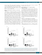
Plasma diminishes loss of δ granule secretion during cold storage
Secretion of granule content in response to PLT activa- tion is required for an efficient hemostatic function. All stored PLTs had a reduced release of granule markers com- pared to freshly isolated ones. However, storage at 4°C improved CD63 release, especially when PLTs were stored in plasma: fold increase of CD63 after activation with TRAP mean±SEM: plasma-APC versus PAS-35-APC, 3.88±0.65 versus 2.71±0.27, respectively, P=0.145 (Figure 4A); and plasma-APC versus PAS-20-APC, 4.75±1.18 versus 2.75±0.75, respectively, P=0.045 (Figure 4B). Storage solu- tion and temperature had only a minor impact on the stor- age lesions causing diminished CD62P expression (Figure 4C and D).
Validation of cold PLT functions in stored APCs
As a step towards optimization of cold storage in rou- tine clinical practice, we next evaluated the effect of cold storage using PAS-35-APCs produced under GMP condi- tions to demonstrate the quality of cold-stored PLTs pre- pared for clinical use throughout extended storage for ten days.
Platelet aggregation in response to collagen was signifi- cantly higher when PLTs were stored at 4°C (Figure 5A). A
AB
CD
Figure 4. The impact of storage temperature and residual plasma concentration on granules release upon activation. After seven days of storage, CD63 (A and B) and CD62P (C and D) expressions were determined for apheresis platelet concentrates (APCs) containing 100% plasma (Plasma-APC), 35% plasma (PAS-35-APC) and 20% plasma (PAS-20-APC) after activation with TRAP. Freshly isolated platelets (PLTs) were used as control for both markers. Data are shown as mean± Standard Error of Mean of fold increase of mean florescence intensity compared to buffer as baseline. *P<0.05. ns: not significant (n=4). RT: room temperature.
haematologica | 2019; 104(1)
211
