
Page 219 - 2019_01-Haematologica-web
P. 219
The impact of plasma on cold storage of APC
Supplementary Figure S1A). In contrast, PLTs stored in PAS with only 20% residual plasma were cleared faster from the mouse circulation compared to those stored in 100% plasma, and this was even more pronounced in cold- stored PLTs (Online Supplementary Figure S1B).
Cold-induced apoptosis rather than desialylation is associated with accelerated clearance of cold-stored platelets, irrespective of residual plasma
Cold storage of PLTs in plasma has been reported to induce desialylation of PLTs. Therefore, we tested the
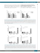
AB
Figure 1. Relative platelet survival after cold storage at different residual plasma concentrations (PC). Apheresis platelet concentrates (APCs) were collected, split and stored at room temperature (RT) or at 4°C, either in 100% plasma (Plasma-APC, black bars) or in platelet additive solution (PAS, gray bars) at two different resid- ual plasma concentrations: (A) 35% (PAS-35-APC) and (B) 20% (PAS-20-APC). Platelets (PLTs) were obtained from APCs at storage day 7 and administered into the mouse circulation via the lateral tail vein. Survival of human PLTs in mouse was analyzed by collecting murine blood after two and five hours. For statistical compar- ison between different groups, the percentage of circulating human PLTs was normalized to Plasma-APC in each corresponding experiment (Plasma-APC was con- sidered 1.0). Data are shown as mean±Standard Error of Mean. *P<0.05; **P<0.01; ***P<0.001; ****P<0.0001. ns: not significant (n=5). Survival curves are available in Online Supplementary Figure S1.
Figure 2. Desialylation and apoptosis after cold storage. Platelet (PLT) desialylation (A and B) and apoptosis (C and D) were analyzed after seven days of storage, at room temperature (RT) or 4°C, in apheresis platelet concentrates (APCs) containing (A and C) 35% plasma (PAS-35-APC), (B and D) 20% plasma (PAS-20-APC) or 100% plasma (Plasma-APC). Desialylation was determined by measurement of FITC-labeled RCA (0.5 mg/mL) that binds to beta (β)-galactose using flow cytometetry. Fresh PLTs incubated with or without neuraminidase (Neu) were used as positive and negative control, respectively. The percentage of apoptotic cells was measured using FITC-labeled Annexin V. As positive (Pos ctr) and negative control (Neg ctr), freshly isolated PLTs incubated in the presence or in the absence of Ionomycin were used. Data are shown as mean±Standard Error of Mean of fluorescence intensity (MFI) and percentage of positive events, respectively. *P<0.05. ns: not significant (n=4).
AB
CD
haematologica | 2019; 104(1)
209
