
Page 222 - 2019_01-Haematologica-web
P. 222
I. Marini et al.
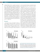
significantly higher response to collagen was found after four days of storage at 4°C in comparison to RT (percent- age of maximal aggregation after collagen, mean±SEM 67±6 vs. 22±5, respectively, P<0.0001), consistent with their better adhesion to collagen (Figure 3). In addition, the δ-granule-dependent response to ADP was reduced less during storage in PAS at 4°C compared to RT (Figure 5B). Ristocetin-mediated PLT agglutination was slightly lower at 4°C compared to RT (Figure 5C). Along the same lines as for PLT aggregation, after activation with TRAP a slightly higher expression of CD62P and CD63 was observed on cold-stored PLTs and the gradual reduction in PLT response was slower (Figure 6A and B). In contrast, no significant impact on the ability of conformational change in the integrin alpha (α)IIb/β (β)III was found during stor- age, regardless of temperature (Figure 6C). Furthermore, similar glucose consumption, and lactate release was found in both storage conditions (Online Supplementary Figure S3A and B, respectively). Finally, although hypoton- ic shock reaction decreased in a time-dependent manner, it was always higher when PLTs were stored at RT com- pared to 4°C (Figure 6D).
Discussion
In the present study, we investigated the impact of residual plasma concentration in APCs on cold storage lesions, including in vivo survival and in vitro hemostatic functions. Despite the reduced in vivo survival of cold- stored PLTs, we showed that cold storage not only pre- serves PLT response to aggregation agonists, but also maintains their adhesion to thrombogenic surfaces better than RT storage. Substituting 65% of plasma with PAS did
not have any important impact on cold-induced storage lesions. However, poor survival and functional results were observed when plasma concentration was further reduced to 20%.
Attempts to store APCs at 4°C were impeded by a short- er survival of cold-stored PLTs. In our study, PLTs from APCs stored at 4°C always showed inferior survival curves compared to those stored at RT. This confirmed previous reports on accelerated clearance of cold-stored PLTs.13,14 Data from an animal model suggested that desialylation of GPIb is responsible for the accelerated elimination of cold- stored PLTs.13,24 However, we found similar β-galactose exposure on RT- and cold-stored PLTs. This fits the results of clinical studies which showed that reconsti- tution of sialylation in human PLTs does not prevent the accelerated clearance of cold-stored APCs.25 Interestingly, we found that cold storage triggers phosphatidylserine exposure indicating higher PLT apoptosis. Although the differences were not always significant, there is an obvious trend. This may indicate that impaired in vivo survival of cold-stored PLTs is actually caused by apoptosis-related mechanisms. In fact, cold storage had previously been found to induce GPIb α-clustering which makes the recep- tors an initiation site for PLT apoptosis and macrophage recognition.26 The inhibition of prostacyclin has been sug- gested to reduce cold-induced changes in GPIb,27 which could be emphasized by our results. Interestingly, cold- induced apoptosis in our study was not accompanied by higher spontaneous expression of P-selectin or CD63, indi- cating that apoptosis and activation during PLT storage are mediated by two independent mechanisms. Therefore, selective targeting of cold-induced apoptosis may provide a new approach to reduce PLT storage lesions. In fact, it was recently reported that cold storage of PLTs enhances
AB
C
Figure 5. Functional analyses of cold-stored apheresis platelet concentrates.
Apheresis platelet concentrates (APCs) were produced under Good Manufacturing Practice. conditions and stored in platelet additive solution (PAS) at a residual plasma concentration of 35% (PAS-35-APC) at room temperature (RT) or 4°C. The maximal aggregation ability of platelets (PLTs) was determined after activation using three inductors: (A) collagen (8 mg/mL), (B) ADP (80 mM) and (C) ristocetin (1.5 mM) (agglutination). Data are shown as mean±Standard Error of Mean. *P<0.05; ****P<0.0001. ns: not significant (n=4).
212
haematologica | 2019; 104(1)
