
Page 177 - 2019_01-Haematologica-web
P. 177
Demographic features such as race, gender and age are shown to associate with disease prevalence and severity. For instance, iTTP occurs more commonly in African- American females11,12 and, perhaps not surprisingly, older age (>60 years) is associated with an increased mortali- ty.12,13 Additionally, serum levels of creatine kinase-mus- cle/brain (CK-MB), troponin I,14 lactate dehydrogenase (LDH), ADAMTS13 antigen or activity levels, anti- ADAMTS13 antibody levels13 and, more recently, the platelet recovery rate15 are shown to be associated with increased mortality.
In this study, we describe the Alabama cohort of 73 unique patients with confirmed diagnosis of iTTP select- ed from a total of 142 admissions. This cohort of patients was primarily from the Southeastern United States. Clinical information, laboratory values, and various bio- markers were collected and analysed with respect to their associations with admission type, disease severity, and mortality.
Methods
Patients
The Institutional Review Board (IRB) of the University of Alabama at Birmingham (UAB) has approved the study protocol. UAB medical center serves as a referral center for the diagnosis and management of patients with thrombotic microangiopathy (TMA) for the state of Alabama and several neighboring states in the Southeast United State of America. Some patients were initially seen by a primary care physician, local internist, or hematologist. If TMA was suspected, patients were referred to the UAB Medical Center for further evaluation and treatment, which may have involved a delay in diagnosis and treatment of one to several days. There were also patients who came directly to the UAB Emergency Department (ED). Within hours of arrival at UAB, a central intravenous catheter was inserted, blood sam- ples were collected for laboratory tests including ADAMTS13 activity and inhibitors, and therapeutic plasma exchange (TPE) was urgently initiated. Seventy-three patients at the UAB Medical Center, between April 2006 and December 2017, were included in this study. Control samples were collected from healthy individuals (age 27-69 years), both male (1/3) and female (2/3), representing the local ethnic population, who did not have a history of hematological diseases, malignancy, and acute inflammatory disorders.
Whole blood was anticoagulated with 3.2% sodium citrate; plasma was separated within four hours of collection, and stored at -80°C prior to analysis. Clinical data pertinent to each patient, including demographic information, past and current medical history, signs and symptoms on admission, laboratory test results, presumptive and final diagnosis, hospital-course, out- come and long-term follow up, were collected by a physician and maintained in the Alabama Registry Database.
Exclusion and inclusion criteria
Patients were excluded from analysis if their final diagnosis was determined to be an alternative TMA, for example: atypical hemolytic uremic syndrome (aHUS), congenital TTP, HIV-relat- ed thrombocytopenia, HELLP syndrome, a life-threatening con- dition during pregnancy with clinical features of hemolysis, ele- vated liver enzymes, and low platelet count,16 TMA following solid organ or hematopoietic stem cell transplantation, drug- induced TTP (i.e., clopidogrel, ticlopidine or gemcitabine) and/or sepsis. Additionally, we excluded patients who were treated
Prognostic markers in autoimmune TTP
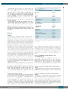
Table 1. Demographics, clinical presentations, and comorbidities in 73 unique patients with iTTP.
Parameters
Age (Years) Sex
Female
Male
Ethnicity
African American Caucasian Afro-hispanic
Presenting symptoms CNS symptoms Abdominal pain Chest pain
Comorbidities
Hypertension
Diabetes mellitus
Systemic lupus erythematosus
Pregnancy
Smoking
Drug use
Values*
41 (32, 52)
41 (56%) 32 (44%)
56 (76.7%) 16 (21.9%) 1 (1.4%)
40 (54.7%) 28 (38.4%) 7 (9.6%)
38 (52.1%)
13 (17.8%)
8 (11.0%)
3 (4.1%)
36 (49.3%)
15 (20.5%)
haematologica | 2019; 104(1)
*All values are expressed as the number and percentage of patients (in parenthesis) in each category except for age, which is expressed as the median and 95% confi- dence interval.
prior to sample drawn with plasma infusion (>3L) and/or TPE and those in remission. Thus, this cohort includes patients expe- riencing their first episode or an exacerbation, or a relapse (only if the sample from the initial episode was not available). Confirmatory tests for ADAMTS13 activity and inhibitors were performed at the Blood Center of Wisconsin (Milwaukee, WI, USA).
Assays for ADAMTS13 activity, inhibitors, and anti-ADAMTS13 IgG
Plasma ADAMTS13 activity and inhibitor titers were deter- mined using a commerical FRETS-VWF73 assay17 and an in- house FRETS-based assay as previously described.18 Plasma anti- ADAMTS13 IgG was determined by an enzyme-linked immunosorbent assay (ELISA) (Diapharma, West Chester, Ohio) in accordance with the manufacturer’s recommendations.
Assays for plasma VWF antigen and collagen-binding activity
Plasma VWF antigen (VWF-Ag) and collagen-binding activity (VWF-CBA) levels were determined using in-house ELISA-based assays as previously described.19
Assays for complement activation and inflammatory markers
Plasma levels of complement activation markers including iC3b, sC5b-9, Bb, and C4d were determined using a commercial ELISA assays (MicroVue, San Diego, CA, USA) following manu- facturer's instructions.8 Plasma HNP1-3 levels were also deter- mined by an ELISA assay, which recognizes all HNP1-3 (Hycult Biotech, Plymouth Meeting, PA).8 Finally, plasma histone-DNA complexes were quantified by the ELISA assay previously described (Roche, Indianapolis, IN, USA).20
167
