
Page 150 - 2019_01-Haematologica-web
P. 150
R. Gressin et al.
formed before treatment (baseline), after four courses of treatment (mid-term MRD), and at the end of treatment (after 6 courses of RiBVD) in peripheral blood and bone marrow until progression or relapse, for a maximum follow-up period of 3 years.18,19 During the follow-up, MRD was evaluated in the blood at 3 monthly intervals for 1 year and every 6 months thereafter while bone marrow MRD monitoring was performed at yearly intervals. A description of additional methods, the MRD study cohort, sample source and numbers is given in the Online Supplementary Methods and illustrat- ed in Online Supplementary Figure S1.
Sample size calculation and statistical analysis
The primary objective of the study was to prolong PFS by 6 months compared to the 18-month median PFS reported for patients treated with R-CHOP-21.3 The number of patients to be enrolled was calculated by a one-step Fleming method. In order to define superiority of the RiBVD regimen over R-CHOP, a PFS rate of 65% or more (H1) was required at 18 months. The treatment was to be considered a failure if the PFS rate at 18 months was ≤50%. Taking into account alpha and beta risks of 5% and 20%, respectively, 69 patients needed to be enrolled. Based on a maxi- mum 10% error in diagnosis, 76 patients had to be enrolled. Additional details are given in the Online Supplementary Methods.
Results
Patients
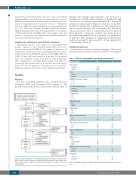
A total of 76 MCL patients were enrolled between November 2011 and December 2012 (Figure 1). All patients were monitored for 3 years after their last cycle of
Figure 1. Consort diagram for the RiBVD phase 2 trial. MCL: mantle cell lym- phoma; DLBCL: diffuse large B-cell lymphoma; HBV: hepatitis B virus; MRD: min- imal residual disease; BM: bone marrow; PML: progressive multifocal leukoen- cephalopathy.
therapy. Two patients were excluded - one because of a misdiagnosis of MCL (diffuse large B-cell lymphoma) and one because of exclusion criteria (hepatitis B) – leaving 74 patients for data analyses (Figure 1). Seventy-one patients had MCL confirmed by central review. The diagnosis was made on tumor biopsies (45 on lymph nodes and 26 on extra-nodal tissue). Due to unsuccessful tissue biopsy in three patients, a diagnosis of MCL was made by flow cytometry in peripheral blood (1 patient) or bone marrow (2 patients). Ki67 staining was performed in 56 patients, and was found ≥30% positive in 59% of these patients (31 of 56 patients) (Table 2).
Treatment response
Seventy-four patients initiated therapy. Sixty-seven patients received at least four cycles (90.5%) of treatment
Table 1. Patients’ demographics and clinical characteristics. Characteristics N. %
Age (years)
median 73
range
64-83
Sex
male 49 66 female 25 34
WHO Performance Status
0-1 73 85 2-4 11 15
Lactate dehydrogenase
normal 44 61 >normal 28 39
B symptoms
no 56 76 yes 17 24
Ann Arbor stage
II 4 6 III-IV 70 94
Bulky tumor
no 52 71 yes 21 29
Extranodal involvement
no 7 9 yes 67 91
Bone marrow involvement
no 24 34 yes 46 66
Spleen involvement
no 38 52 yes 35 48
MIPI score
low 2 3 intermediate 12 17 high 58 80
MIB1 / ki67 prolferation index
<30% 21 41 ≥30% 30 59
Pathology
classic 61 86
blastoid 10 14
WHO:World Heath Organization; MIPI:Mantle Cell Lymphoma International Prognostic Index.
140
haematologica | 2019; 104(1)
