
Page 145 - 2019_01-Haematologica-web
P. 145
Fusion genes involving MEF2D in B-ALL
As TCF3-PBX1-positive cases also express the cytoplas- mic m chain-positive pre-B ALL immunophenotype,28,29 we subsequently compared the gene expression of MEF2D fusion cases with TCF3-PBX1-positive cases as well as B- other-ALL. As shown in Online Supplementary Figure S4, MEF2D fusion patients were clearly separate, within a dis- tinct cluster from TCF3-PBX1-positive and B-other-ALL cases, based on unsupervised hierarchical clustering. Up- regulated genes common to both MEF2D fusion and TCF3-PBX1-positive cases compared to B-other-ALL included: IKZF2, IRF4, TCL6, IGHG, IGHV5-78, IGLL1, VPREB3, and BCL2L11, while RUNX2, IRF9, CRLF2, CD34, and CCND2 were common down-regulated genes (summarized in Online Supplementary Figure S5). HDAC9 was also identified as a common up-regulated gene in both MEF2D fusion and TCF3-PBX1-positive cases, whereas its expression was significantly higher in MEF2D fusion than TCF3-PBX1-positive cases. On the other hand, MEF2D, MME (coding CD10), and RAG1 were down-reg- ulated in MEF2D fusion, but not in TCF3-PBX1-positive cases, thus the low-level expression of CD10 seen in MEF2D fusion cases was identified at the gene-expression level. Interestingly, GATA3 was identified as a highly expressed gene in MEF2D fusion cases, yet it was signifi- cantly down-regulated in TCF3-PBX1-positive cases.
To explore the gene expression characteristics of MEF2D fusion ALL connected with B-cell differentiation, we performed GSEA using 18 curated gene sets of B lym- phocytes at various differentiation stages, as well as 7 early hematopoietic stages including stem cells. Firstly, we compared the gene expression signatures of B-ALL cases with different types of genetic abnormalities. Using B- other-ALL as a reference control, we observed that the majority of gene expression signatures found in B lympho- cytes at various differentiation stages were enriched in ALL cases with TCF3-PBX1 (Online Supplementary Table S10 and S11). In contrast, only three gene sets were enriched in MEF2D fusion ALL. We further examined the gene expression characteristics of MEF2D fusion ALL by direct comparison with TCF3-PBX1, and observed that most of the signatures of differentiation stage-specific B lymphocytes as well as early hematopoietic populations were enriched in TCF3-PBX1, but not MEF2D fusion ALL (Online Supplementary Table S10).
Clinical characteristics and outcomes of MEF2D fusion-positive patients
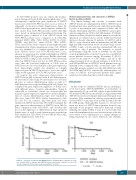
The clinical findings and outcome of patients with MEF2D fusions are summarized in Table 1. MEF2D fusion B-ALL patients were aged between 3 and 15 years (median: 9 years) at presentation and comprised 6 males and 10 females. Their initial white blood cell (WBC) counts at pres- entation ranged from 3,400 to 124,100 (median: 27,300/ml). Analysis of fluids obtained by lumbar puncture revealed no indication of central nervous system (CNS) involvement (data not shown). Among 13 patients, 10 (62.5%) and 4 (25.0%) were classified with an intermediate risk (IR) and high risk (HR), respectively, based on advanced age, elevat- ed WBC counts, or both, and thus standard risk (SR) was assigned to only 2 patients. The response to steroid monotherapy, using the cutoff of 1,000/mL for the blast count in peripheral blood on day 8 was not poor in MEF2D fusion patients; however, among 15 patients, 8 (53.3%) showed bone marrow or CNS relapse, and all of the relapsed patients died. As shown in Figure 4, both the 5- year EFS and OS rates for MEF2D fusion patients were sig- nificantly lower compared with a consecutive series of B- other-ALL patients enrolled onto the L04-16/L06-16 study (P=0.0306 and P=0.0013, respectively), indicating that out- comes for MEF2D fusion-positive patients were signifi- cantly less favorable than those with B-other-ALL.
Discussion
In this study, we identified MEF2D fusions, including a novel fusion gene, MEF2D-HNRNPH1, at an incidence of approximately 2% in our B-ALL cohort. It was notable that we identified MEF2D fusions in B-LBL. B-ALL patients with MEF2D fusions showed unique clinical and biological char- acteristics. They exhibited an older age at presentation and elevated WBC counts, thus were mostly classified into the IR or HR groups. Although their response to steroid treat- ment was not poor, MEF2D fusion patients showed a sig- nificantly worse prognosis with more than half of them relapsing and dying within 1 year. It is noteworthy that stem cell transplantation was not effective in any of the five cases where it was administered as a salvage therapy for relapsed patients in our cohort. Therefore, the establish-
Figure 4. Outcomes of patients with MEF2D fusions. (A) Kaplan-Meier estimates of event- free survival (EFS) of patients with MEF2D fusions, and B-other, log-rank P=0.306). (B) Overall survival (OS) for the same as above (log-rank P=0.0013).
P=0.0306 P=0.0013
haematologica | 2019; 104(1)
135
