
Page 142 - 2019_01-Haematologica-web
P. 142
K. Ohki et al.
well as the known MEF2D fusions, we identified MEF2D- HNRNPH1 as a novel fusion in 1 patient (Figure 1, Table 1, Case 17). Among the L04-16/L06-16 cohort,11,19 comprising a consecutive series of 290 B-ALL patients, including 126 classified as B-other-ALL, 5 MEF2D-BCL9 and 2 MEF2D- HNRNPUL1 patients were identified (Online Supplementary Table S1 and Figure S1). The incidence of MEF2D fusions in childhood ALL, calculated from this
cohort, was 5.6% in B-other-ALL and 2.4% in B-ALL over- all. MEF2D-BCL9 was the most recurrent, at a frequency of 4.0% in B-other-ALL and 1.7% in B-ALL overall.
Structure of MEF2D fusions
The structure and sequences of MEF2D-BCL9 as well as
a schematic representation of the predicted fusion pro- teins, are depicted in Figure 1, Table 1 and Online
A
B
P<0.01 P<0.05
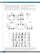
Figure 2. Immunophenotypic characteristics of B-ALL patients with MEF2D fusions. (A) The positivity (percentage) of CD10, cytoplasmic m, CD5, CD38, and CD33 of MEF2D fusion-positive, TCF3-PBX1-positive, and B-other patients are plotted as a scattergram. A detailed list of positivity for each immunophenotypic marker of the patients is presented in Online Supplementary Table S4. (B) Typical histograms of CD10, CD19, cytoplasmic m, aberrant CD5, CD33, and CD38 of MEF2D-BCL9, MEF2D-HNRNPUL1, TCF3-PBX1, and B-other patients are indicated with a positive rate (%). X-axis, fluorescence intensity; Y-axis, relative cell number.
132
haematologica | 2019; 104(1)
