
Page 152 - 2018_12-Haematologica-web
P. 152
L.V. Abruzzo et al.
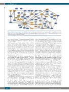
Figure 3. Construction of a specific trisomy 12 (+12) CLL gene expression network. Genes indicated in blue are over-expressed in +12 chronic lymphocytic leukemia compared to other cytogenetic subtypes. Genes indicated in orange are under-expressed in +12 CLL. Genes indicated in gray are not differentially expressed. Brighter colors are more statistically significant; duller colors are less statistically significant. Genes on chromosome 12 are indicated by hexagons; genes located on other chromosomes are indicated by rectangles.
due to greater reliance on microenvironmental survival signals, another factor that may contribute to their good response to FCR.30
We identified three NFAT mRNAs, NFATc1 (NFAT2), NFATc2 (NFAT1), and NFATc3 (NFAT4), that are over- expressed in +12 CLL, and involved in three activated sig- naling pathways: protein kinase A, phospholipase C, and integrin signaling. The NFAT (nuclear factor of activated T cells) transcription factor family contains five proteins; four are regulated by calcium and the calcineurin signaling pathway.34,35 Originally described in T cells, NFAT proteins are expressed by B cells, natural killer cells, and other cell types. They regulate genes involved in cell cycle, apopto- sis, angiogenesis, and metastasis.
In resting lymphocytes, inactive hyperphosphorylated NFAT proteins are confined to the cytoplasm.34,35 In B cells, BCR ligation by cognate antigen activates SYK, which phosphorylates and activates BTK.36 BTK then phospho- rylates and activates PLCg2, which catalyzes the hydroly- sis of inositol 1, 4, 5-trisphosphate (IP3) and diacylglycerol (DAG). IP3 mediates influx of extracellular calcium and calcium release from intracellular stores, which results in calcium/calmodulin-dependent activation of calcineurin. In the cytoplasm, calcineurin cleaves phosphate groups from inactive, hyperphosphorylated NFAT proteins, which enter the nucleus, bind to specific response ele- ments in target gene promoters (alone or in combination with partner proteins), and activate or inhibit transcrip- tion. Thus, NFAT proteins integrate calcium signaling with other signaling pathways, including the MAPKinase, WNT, and NOTCH pathways.
Dysregulated calcineurin/NFAT signaling has been reported in carcinomas and lymphoid malignancies. In large B-cell lymphomas, active NFAT interacts with NF-kB and directly regulates CD154 expression to maintain growth.37 Despite its central role in BCR signaling, there are few studies of NFAT signaling in CLL.38-41 LeRoy et al. demonstrated that BCR-NFAT signaling affects CLL clini- cal outcome, and suggest that BCR-NFAT intermediates
serve as therapeutic targets.38 Recently, Oakes et al. com- pared the epigenetic programming of normal B-cell sub- sets with 268 CLL samples, and identified aberrant NFAT methylation in a CLL subset.42 Thus, efforts are underway to develop more specific NFAT inhibitors.43,44
Using IPA to construct a novel +12 specific network, we identified ecto-5’-nucleotidase (NT5E, CD73) as an impor- tant element. NT5E, an immune checkpoint molecule of potential therapeutic value, is expressed in a wide variety of tissues.45-47 Immune checkpoint molecules regulate interac- tions between immune and tumor cells, and may stimulate or inhibit these interactions.45,46 Many cancers exploit these molecules to evade an anti-tumor immune response. Among the best described are the inhibitory molecules PD1, PD-L1, and CTLA-4. CLL is characterized by immunosuppression and an inefficient anti-tumor response that results from defects in humoral and cellular immunity, including ineffective T-cell responses and expression of exhaustion-like surface markers, such as PD-L1.48-50 Immune checkpoint inhibitors that target the PD1/PD-L1 and CTLA- 4 pathways are being used to treat a variety of tumors, including melanoma and prostate cancer.
NT5E catalyzes the conversion of extracellular ATP to adenosine, which is critical for immune function.45,46 Among immune cells, it is expressed in macrophages, B cells, regulatory T cells, and dendritic cells. NT5E helps tumors evade the immune response by inhibiting the acti- vation, proliferation, and homing of tumor-specific T cells, and by enhancing conversion of anti-tumor type 1 macrophages to pro-tumor type 2 macrophages. The NT5E-adenosine axis constitutes a promising new path- way in cancer immunotherapy. Targeted blockade of NT5E or adenosine receptors promotes anti-tumor immu- nity and enhances the activity of first-generation immune checkpoint blockers.45,46 Phase I clinical trials evaluating the efficacy of anti-NT5E or anti-A2A therapies in cancer patients are underway. However, few studies have inves- tigated the functions of NT5E in lymphoid malignancies. In a study of CLL patients, 30% of cases expressed NT5E,
2076
haematologica | 2018; 103(12)
