
Page 140 - 2018_12-Haematologica-web
P. 140
S. Kinoshita et al.
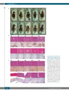
A
B
C
Figure 4. Macroscopic and microscop- ic findings in primary ANKL cell-bear- ing mice treated or not treated with BAY 1143572. (A) Macroscopic appearance of control mice treated with vehicle (control; top panels) or BAY 1143572 (bottom panels). Liver and spleen are demarcated by narrow yellow lines. (B) Photomicrographs of control mouse liver (hematoxylin and eosin [HE] staining) (top panels), and with in situ hybridization (ISH) using an Epstein–Barr virus (EBV)-encoded RNA (EBER) probe (panels second from top). HE-stained liver (third panels from the top), together with ISH using an EBER Probe (bottom panels) from BAY 1143572-treated mice. Scale bars represent 100 mm. (C) HE stain- ing of the spleen of control mice (top panels), together with ISH using an EBER Probe (Leica Microsystems Newcastle Ltd.) (second panels from the top). HE staining of the spleen (third panels from the top), and ISH using an EBER Probe (bottom panels) from mice treated with BAY 1143572. Scale bars represent 100 mm.
2064
haematologica | 2018; 103(12)
