
Page 138 - 2018_12-Haematologica-web
P. 138
S. Kinoshita et al.
sure to 100.0 uM BAY 1143572 for 24 hours (Figure 3A). On the other hand, IC50 values for BAY 1143572 after 24 hours of incubation with CD56-positive tumor cells obtained from two different patients with ANKL were 0.16 uM and 0.29 uM (Figure 3B). In contrast to the CD56-positive cells from healthy volunteers, most of the tumor cells were dead after exposure to 3.0 uM BAY 1143572 for 24 hours. Bay 1143572 inhibited phosphory- lation of RNAPII CTD at the Ser2 site and reduced the expression of Mcl-1 proteins in the primary tumor cells (Figure 3C).
Phosphorylation status of RNAPII at the Ser2 site in tumor lesions of ENKTL, nasal type
RNAPII in ENKTL, nasal type, tumor cells were highly phosphorylated at Ser2 sites. This was also the case in non-tumorous lymphocytes from reactive lymph nodes. Indeed, the degree of phosphorylation of RNAPII at the Ser2 site was identical in both the tumor cells and non- tumor lymphocytes. Three representative cases from each group are shown in Online Supplementary Figure S2.
Macroscopic and microscopic findings in primary ANKL cell-bearing mice with or without BAY 1143572 therapy
The appearance of mice treated with vehicle only and those treated with BAY 1143572, 22 days after primary ANKL cell inoculation, is shown in Figure 4A, upper and lower panels, respectively. Hepatosplenomegaly was observed in all of the untreated mice, but not in those treated with BAY 1143572 (liver and spleen are demarcat- ed by narrow yellow lines). Immunohistological analyses revealed that the livers of the control mice were infiltrated by atypical lymphoid cells, and the normal architecture was destroyed (Figure 4B, top panels). ISH showed that these atypical cells were positive for EBER (Figure 4B, sec- ond panels from the top). On the other hand, the liver architecture of the treated mice was almost intact (Figure 4B, third panels from the top), and EBER-positive cells
were rare (Figure 4B, bottom panels). The same immuno- histological analyses also revealed that the spleens of the untreated mice were infiltrated by atypical lymphoid cells, and the normal architecture was completely destroyed (Figure 4C, top panels). ISH also showed that these atyp- ical cells were positive for EBER (Figure 4C, second panels from the top). On the other hand, the splenic architecture of the treated mice, like the liver, was essentially intact (Figure 4C, third panels from the top), and EBER-positive cells were rarely observed (Figure 4C, bottom panels).
BAY 1143572 treatment reduces primary ANKL cells in the blood of mice
Twenty-two days after inoculation of primary ANKL cells, the percentage of human tumor cells (identified as CD45- and CD16/56-positive but CD19-negative) in the whole blood of control NOG mouse No.1 was 17.1% (i.e., 17.3% [CD45-positive lymphocyte population] x 98.7% [CD16/56-positive, but CD19-negative cells] = 17.1%) (Figure 5A, the two upper left panels). In control NOG mice Nos. 2, 3, 4, and 5, and in BAY1143572-treated NOG mice Nos. 1, 2, 3, 4, and 5, the percentages of ANKL cells in whole blood, calculated in the same manner, were 30.9, 45.0, 11.1, and 38.4%; and 0.6, 0.1, 0.4, 0.4, and 0.4%, respectively. Thus, BAY 1143572 treatment resulted in significantly decreased percentages of ANKL cells in the blood of these xenogeneic primary tumor-bearing mice (P=0.009; Figure 5A).
BAY 1143572 treatment reduces primary ANKL cells in the bone marrow
Twenty-two days after inoculation, the percentage of ANKL tumor cells in the bone marrow of control NOG mouse No.1 was 18.3% (i.e., 26.5% [CD45-positive lym- phocyte population] x 69.2% [CD16/56-positive, CD19- negative] = 18.3%) (Figure 8, the two upper left panels). In control NOG mice Nos. 2, 3, 4, and 5, and in BAY 1143572-treated NOG mice Nos. 1, 2, 3, 4, and 5, the per- centages of ANKL cells in bone marrow, calculated in the
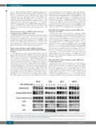
Figure 2. BAY 1143572 affects CDK9 activity in NK-cell leukemia/lymphoma lines. NK-92, MTA, KAI-3, and KHYG-1 cells were treated with the indicated concen- trations of BAY 1143572 for 5 hours, followed by Western blotting probing RNA polymerase II (RNAPII), phospho-RNAPII (the serine-2 residue of the C-terminal domain [CTD] [Ser2]), phospho-RNAPII (Ser5), c-Myc, and Mcl-1. Actin was the loading control.
2062
haematologica | 2018; 103(12)
