
Page 27 - 2018_11-Haematologica-web
P. 27
Preclinical models of mastocytosis
clones.86 However, the mTOR inhibitor rapamycin induced apoptosis only in HMC-1.2 cells.86 To support this unexpected selectivity, the authors demonstrated that rapamycin inhibited the phosphorylation of 4E-BP1, a downstream substrate of the mTOR pathway, only in HMC-1.2 cells.86 More recently, it was reported that the dual PI3-kinase/mTOR blocker NVP-BEZ235 has similar growth inhibitory effects in HMC-1.1 and HMC-1.2 cells.87 However, despite these encouraging data, no objective response was observed in a study in which everolimus, an oral mTOR inhibitor, was given at a dose of 10 mg daily to ten SM patients.88
Finally, aberrant accumulation of neoplastic MC in SM might result from deregulation of apoptosis pathways.89 Indeed, the anti-apoptotic molecules BCL-2, BCL-xL and MCL-1 are overexpressed in KIT D816V+ neoplastic MC in SM patients,90-92 while the expression of the pro-apop- totic molecule BIM is suppressed in these cells.93 It has also been reported that MCL-1 is detectable in HMC-1.1 and HMC-1.2 cells.93 Moreover, exposure of these cells to MCL-1-specific antisense oligonucleotides or to MCL-1- specific short interfering RNA resulted in reduced cell sur- vival and increased apoptosis.93 In further studies, evi- dence was provided that the pan-BCL-2 family blocker obatoclax inhibited the proliferation of HMC-1 cells, together with increased expression of PUMA, NOXA, and BIM mRNA, and apoptosis.94
Human mastocytosis-like mast cell lines and drugs targeting surface antigens or epigenetic regulators
Although drugs targeting KIT D816V have demonstrat- ed activity on MC in vitro and in vivo, these agents do not cure patients with advanced SM.48,49,71 Apart from several different mechanisms of resistance, neoplastic cells in these patients may exhibit a complex pattern of genetic alterations together with, or even often preceding the appearance of, the KIT mutant, as it is the case for TET2, SRSF2 and ASXL1 mutants,37 which could explain resist- ance to TKI. For this reason, attention has been focused recently on alternative targets which could help to over- come such resistance, namely surface antigens specifical- ly or aberrantly expressed by neoplastic MC and epige-
netic targets. Antibodies or drugs directed against these targets may also be able to overcome intrinsic neoplastic stem cell resistance, often associated with quiescence and altered drug influx or rapid drug efflux. Antibody-based drugs may exert antineoplastic effects independently of such mechanisms of resistance.
Targeting surface antigens
Several antigens are aberrantly expressed or overex- pressed on neoplastic MC and on their progenitors in SM, including CD13, CD25, CD30, CD33, CD44, CD52, CD117 and CD123,95-97 and might, therefore, be consid- ered as potential therapeutic targets. Table 4 provides an overview of cell surface targets expressed on human MCL-like cell lines.
CD30 is aberrantly expressed by neoplastic MC in a subset of patients with SM, but not by normal/reactive MC.98 In a recent study, it was observed that the MCPV- 1.1 subclone expressed high levels of CD30, while HMC- 1.1 cells expressed low CD30 levels, and HMC-1.2 cells did not express CD30.99 The CD30-targeting antibody- conjugate brentuximab-vedotin inhibited the in vitro pro- liferation of neoplastic MC, with lower IC50 values obtained for MCPV-1.1 cells (10 mg/mL) than for HMC- 1.2 cells (>50 mg/mL).99 In addition, brentuximab-vedotin produced apoptosis in primary CD30+ neoplastic MC.99 However, overall, the effects of brentuximab-vedotin on neoplastic MC are relatively weak. Correspondingly, no major clinical activity has been reported in clinical trials to date. In addition, neoplastic stem cells in advanced SM usually lack CD30 (personal information, PV).
In contrast to normal MC and MC from ISM patients, CD52 is abundantly expressed on neoplastic MC in most patients with advanced SM.16 Recently, it was reported that the CD52-targeting antibody alemtuzumab counter- acts growth of MCPV-1.1 cells.16 Additionally, MCPV-1.1 cells were injected into NSG mice which were then treat- ed with alemtuzumab or control vehicle. The alem- tuzumab-treated mice had increased survival compared to controls, and reduced organ infiltration by neoplastic MC.16 Given that neoplastic (leukemic) stem cells identi- fied in advanced SM may also express CD52,100 it can be
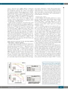
Figure 3. Dose-dependent inhibition of the proliferation of KIT D816V+ human mast cell lines by STAT5 inhibitors in vitro. ROSAKIT D816V and HMC-1.2 cells were cultured in 96-well plates for 72 h in control medium containing 0.1% dimethylsulfoxide (DMSO) or with increasing con- centrations (between 1.0 and 50.0 mM) of SF-1066, a very weak STA5 inhibitor (Ki >25 mM on STAT5), and of more specific and potent STAT5 inhibitors BP-1-102 and BP-1-108 (Ki >10 mM on STAT5).74 Viability was calculated in each condition by the MTT method. Results are expressed as percent of control and represent the mean ± standard deviation of triplicate experiments. The half maximal inhibitory concentration (IC50) at day 3 of each compound for each cell line was calculated using Prism GraphPad 4.0 software after plotting log concentration versus response. As expected, while the IC50 for SF-1066 was >50 mM, IC50 values for the more STAT5-specific com- pounds were lower: 11 mM for BP-1-102 on both KIT D816V+ neoplastic human MC lines and 34 and 22 mM for BP-1-108 for, respectively, HMC-1.2 and ROSAKIT D816V cells. Although these values are still irrelevant at the pharmacological level, they open hopes that drug opti- mization might lead in a near future to the design of more potent small molecules inhibiting STAT5 activity.
haematologica | 2018; 103(11)
1767
