
Page 26 - 2018_11-Haematologica-web
P. 26
M. Arock et al.
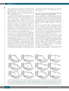
clinical trials have been conducted to determine the effi- cacy of midostaurin in patients with advanced SM, with promising results.48,49,71 An overview of the effects of midostaurin and of several other TKI on the growth of HMC-1 and ROSA cells is presented in Figure 2 and Online Supplementary Table S4.
Several other TKI with different mechanisms of action were also found to exert antineoplastic effects in vitro on KIT D816V+ neoplastic MC, including HMC-1 cells. These drugs include 17-AAG (17-allylamino-17- demethoxygeldanamycin),72 EXEL-0862 (a TKI active against fibroblast growth factor receptors, vascular endothelial growth factor receptors, platelet-derived growth factor receptors, FLT3 and KIT),73 triptolide (a diterpenoid),74 ponatinib (a multi-kinase blocker),68 and bosutinib (a LYN/BTK-inhibiting TKI),28 which was administered to a patient with advanced SM, with no benefit.75 Nilotinib, which showed some effects in vitro on mutant KIT,76 was recently administered to 61 SM patients, with transient activity in some patients.77
More recently, several new KIT-TKI with inhibitory activity in vitro on several KIT mutants, including KIT D816V, have been developed. DCC-2618 (Deciphera Inc.) is a switch control type II KIT inhibitor, which arrests KIT in an inactive state, regardless of activating mutations, such as KIT D816V.78 In a recent study, it was found that DCC-2618 inhibits proliferation and survival of HMC- 1.1, HMC-1.2 and ROSAKIT D816V cells at IC50 <1 mM.79 BLU- 285 is a TKI developed by Blueprint Medicines. At low concentrations, BLU-285 selectively inhibited KIT D816V enzymatic activity (IC50 = 0.27 nM).80 The cellular activity of BLU-285 on this mutant was also measured by autophosphorylation in HMC-1.2 cells with an IC50 of 4.0 nM.80 Finally, BLU-285 inhibited in vitro the proliferation
of KIT D816V+ HMC-1.2 cells with an IC50 of 125 nM, while being less active on KIT D816V– HMC-1.1 cells (IC50 = 344 nM).81
Human mastocytosis-like mast cell lines and drugs targeting KIT- dependent or KIT-independent signaling pathways
Since KIT D816V is equally present in ISM and advanced SM patients, who have different life expectan- cies,82 the current assumption is that additional, KIT-inde- pendent pathways and pro-oncogenic hits and lesions are responsible for disease progression in advanced SM. Such pathways and pro-oncogenic molecules include LYN, BTK, STAT5, PI3-K, mTOR and members of the BCL-2 family.
For instance, LYN and BTK are phosphorylated in HMC-1.1 and HMC-1.2 subclones independently of KIT, and short interfering RNA against LYN and BTK decreased the survival of both subclones.28 In the same set of experiments, dasatinib blocked not only the kinase activity of KIT, but also LYN and BTK activation in neo- plastic MC, while bosutinib inhibited LYN and BTK acti- vation without decreasing KIT kinase activity.28
Another molecule, STAT5, seems critical for KIT D816V-driven proliferation in MCL-like MC lines as well as in neoplastic MC in SM patients.24,83 Chaix et al. report- ed that the KIT D816V mutant can directly phosphorylate STAT5 in vitro.83 Interestingly, STAT5 is transcriptionally active in the HMC-1 cell line and in ROSAKIT D816V cells,15,24 and drugs targeting STAT5 are effective in decreasing the growth rate of these cells.84,85 Figure 3 shows representa- tive curves of dose-dependent inhibition of the viability of MC lines by STAT5 inhibitors.
Gabillot-Carre et al. reported constitutive activation of the mTOR signaling pathway in both HMC-1 sub-
Figure 2. Dose-dependent inhibition of the proliferation of wild-type and mutant KIT human mast cell lines by various tyrosine kinase inhibitors in vitro. Human mast cell lines expressing KIT D816V (HMC-1.2 or ROSAKIT D816V; black lines) or lacking KIT D816V (HMC-1.1 or ROSAKIT WT; gray lines) were incubated in control medium (Co) or in medium containing various concentrations of tyrosine kinase inhibitors (as indicated) at 37°C for 48 h. Thereafter, 3H-thymidine uptake was measured. Results are expressed as percent of 3H-thymidine uptake compared to the control and represent the mean ± standard deviation of three different experiments.
1766
haematologica | 2018; 103(11)
