
Page 65 - 2018_10-Haematologica-web
P. 65
IO impairs normal HSPCs and survival in MDS mice
induce apoptosis of BM cells in normal mice (Figure 5A), which has previously been confirmed by others.14 Of note, normal (GFP–) BM cells in RX291/FE mice have a markedly higher apoptosis level than those of RX291/NS mice, but not mutant (GFP+) cells (Figure 5A), implying that iron overload can promote apoptosis of normal BM
cells in MDS. It has been reported that ROS can induce apoptosis.14 Our data showed that the ROS level was also clearly higher in RX291/FE mice than that of RX291/NS mice (Figure 5B) in BM. And erythroid cells in BM presented similar changes (Figure 5C). We also detected the mRNA levels of NOX4 related to ROS gen-
ABC
DE
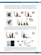
Figure 3. Iron overload impairs the frequency of normal hematopoietic stem and progenitor cells (HSPCs) and affects erythroid maturation in myelodysplastic syn- drome (MDS) mice. (A) Gating hematopoietic stem cells (HSCs) and hematopoietic progenitor cells (HPCs) by flow cytometry. (B) The frequency of HSCs in different groups. (C) Frequency of HPCs in different groups. (D) Different stages of erythroid through CD71 and Ter119 gating. (E) Effect of iron overload on erythroid differ- entiation. *P<0.05, **P<0.01. BMMNCs: bone marrow mononuclear cells; GFP-: normal; GFP+: mutant; LSK: Lin–c-Kit+Sca1+, HSCs; LK: Lin–c-Kit+Sca1–, HPCs; E1: proerythroblasts; E2: basophilic erythroblasts; E3: polychromatophilic erythroblasts; E4: orthochromatic erythroblasts.
Figure 4. Iron overload compromises the erythroid colony-forming capacity of normal hematopoietic stem and progenitor cells (HSPCs) in myelodysplastic syn- drome mice. (A) The colonies generated by GFP- cells. (B) A serial replating assay about GFP– HSPCs. (C) Comparison of the colony-forming capacity between GFP– and GFP+ cells. Left column is in bright field; right column is in fluorescence field. Red arrow indicates the location of GFP+ cells. (D) Experimental scheme of our mouse model using GFP+ cells to perform competitive repopulation assay and detecting chimerism of GFP+ cells post transplantation. *P<0.05. CFU-E: colony-form- ing unit erythroid; BFU-E: burst-forming unit erythroid; GM: colony-forming unit granulocyte-macrophage; GEMM: colony-forming unit granulocyte, erythroid, macrophage, megakarycyte.
AB
CD
haematologica | 2018; 103(10)
1631
