
Page 64 - 2018_10-Haematologica-web
P. 64
X. Jin et al.
matophilic erythroblasts and orthochromatic erythrob- lasts as E1, E2, E3 and E4, respectively. Compared to Empty mice, MDS mice showed a significant increase in the number of GFP- E1 and a significant decrease in GFP- E2 (Figure 3E), which may due to erythroid dysmaturity in MDS. Of note, iron overload can significantly reduce the frequency of E3 and E4 in control mice and GFP– E1 in MDS mice, while it did not exhibit any significant inhibi- tion in GFP+ MDS erythroid cells, which may suggest that iron overload has a negative effect on normal hematopoiesis in erythroid, but not malignant clones. Collectively, our study shows that iron overload impairs the frequency of normal HSPCs and may inhibit erythroid maturation in MDS mice.
Iron overload compromises the erythroid colony-forming capacity of normal HSPCs in MDS mice
We next wanted to examine if iron overload has a neg- ative impact on HSPCs function in MDS mice. Therefore, we collected BM cells and determined the colony-forming capacity or hematopoietic cells reconstitution ability of these cells. Our data showed that normal (GFP–) HSPCs in MDS mice have a significantly lower ability to form colony-forming unit erythroid (CFU-E), burst-forming unit erythroid (BFU-E), and colony-forming unit granulocyte, erythroid, macrophage, megakarycyte (CFU-GEMM) colonies compared to control mice (Figure 4A). What is more, iron overload can markedly inhibit CFU-E and BFU- E of GFP- HSPCs in both control and MDS mice. We also tested a serial replating assay about GFP– HSPCs (Figure 4B). Despite the fact that the first plating did not show sig-
nificantly reduced clonogenic numbers, but there seemed to be a declining trend between Empty/NS and Empty/FE mice, the count of clonogenic HSPCs exhibited a marked decline at the second and subsequent platings (Figure 4B), which supported the view that the colony-forming capac- ity of normal HSPCs was suppressed by iron overload in MDS mice. Because GFP+ HSPCs were unable to form colonies but clusters compared to GFP– HSPCs (Figure 4C), we performed competitive repopulation assay to identify the effect of iron overload on the hematopoietic reconsti- tution capacity of GFP+ cells (Figure 4D). To our surprise, the chimerism of GFP+ cells from RX291/FE mice was not significant compared to those of RX291/NS mice (Figure 4D), indicating that iron overload may not damage the hematopoietic reconstitution capacity of GFP+ HSPCs. Together, we conclude that iron overload can compromise the erythroid colony-forming capacity of normal HSPCs, but may not affect the hematopoietic reconstitution capacity of abnormal HSPCs in MDS.
Iron overload inhibits erythroid hematopoiesis in MDS mice through ROS
These data showed that iron overload inhibits ery- throid hematopoiesis in MDS mice. We next investigated the mechanisms involved in this process. Previous stud- ies have reported that enhanced apoptosis of BM cells in MDS can cause pancytopenia.7 Indeed, our data showed that the level of apoptosis in RX291/NS and RX291/FE mice was significantly increased compared to Empty/NS and Empty/FE group (Figure 5A), respectively, consistent with previous studies.8 In addition, iron overload can
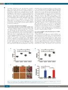
AB
CD
Figure 2. Iron overload model can be established in RUNX1S291fs induced myelodysplastic syndrome mice. (A) Weight of liver. (B) Weight of spleen. (C) Perl’s iron staining of liver, spleen and bone marrow (BM). (D) Ferritin level detected by ELISA assay. **P<0.01, ***P<0.001.
1630
haematologica | 2018; 103(10)
