
Page 67 - 2018_10-Haematologica-web
P. 67
IO impairs normal HSPCs and survival in MDS mice
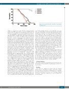
Figure 6. Iron overload shortens the survival time of myelodysplas- tic syndrome mice.
similar to our previous study.13 We also obtained similar results in the PB between the two groups of MDS mice. In addition, we found that iron overload impairs the frequen- cy of normal HSPCs, especially in erythroid cells, but not abnormal HSPCs in MDS mice. We further investigated the impact of iron overload on HSPCs function and found that iron overload compromises the erythroid colony- forming capacity of normal HSPCs, but may not affect the hematopoietic reconstitution capacity of abnormal HSPCs in MDS mice. In addition, RX291/FE mice have a shorter median survival than RX291/NS mice, indicating that iron overload can shorten survival time in MDS mice. There are increasing data to show that oxidative stress was increased in BM cells of patients with iron overload, and antioxidant or iron chelator therapy could partially rescue the impaired hematopoietic function of patients, which indicates the presence of ROS-induced cellular injury.19,23 Of interest, we detected the level of ROS and found it sig- nificantly increased in RX291/FE compared with RX291/NS group, suggesting that iron overload impairs normal HSPCs, at least in part, via inducing ROS in MDS, consistent with previous studies.19,24 Our data showed that iron overload decreased the number of HSPCs partially due to ROS-induced apoptosis. However, whether the reduced HSPCs was regulated by HIF-1a/ROS or NF-κB pathway warrants further investigation.20,24 It has been reported that the TGF-b pathway is myelosuppressive and inhibits erythroid differentiation by induction of ROS and apoptosis in erythroblasts.15-17,25 Our data showed in GDF11, one of the TGF-b superfamilies, mRNA and pro- tein levels significantly increased in RX291/FE mice com- pared to RX291/NS mice, suggesting that iron overload can damage erythroid hematopoiesis in MDS mice, which may partially be due to GDF11-induced ROS, leading to enhanced apoptosis of normal BM cells and inhibition of their function in MDS. However, additional studies are needed to clarify whether GDF11-induced ROS and apop- tosis of erythroid was related to the Fas-Fas ligand path-
way.16,25 Interestingly, Masayo et al. found that iron over- load can activate glucose metabolism and increase DNA methylation, which is associated with MDS pathogenesis and progression, and iron chelation can reverse these effects.26 Therefore, future studies should be conducted to evaluate the impact of iron overload on metabolic path- ways such as glucose and lipid involved in MDS patho- genesis in our model. In addition, our mice can be used to illustrate the association between oxidative imbalance and iron overload in MDS.18 Our previous study showed that damaged mesenchymal stromal cells (MSC) were related to iron overload induced ROS in normal mice.21 However, Zheng et al. reported that iron overload damages MSC through AMPK/MFF/Drp1 pathway in MDS.27 Thus, fur- ther research should investigate the effect of iron over- load on the BM microenvironment in MDS.
In conclusion, our preliminary findings suggest that iron overload impairs the frequency and function of normal HSPCs, particularly in erythroid, at least in part via GDF11-induced ROS, and shortens survival in MDS. Given that there are a few MDS models available, and we are the first to utilize RUNX1-S291fs-induced MDS mice to successfully construct an iron overload model, we hope this model will be helpful for further exploring the influ- ence and mechanism of iron overload on MDS.
Acknowledgments
The authors would like to thank Dr Atsushi Iwam for the valu- able plasmids. We also thank Dr Gang Huang for excellent tech- nical assistance.
Funding
This work was supported by grants from the National Natural Sciences Foundation of China (81400092), Tianjin Key Natural Science Foundation (17JCZDJC35800, 15JCQN- JC45500), and Tianjin Key Science and Technology Program (2015K215, 15KG134, 16KG110), as well as Tianjin First Central Hospital.
haematologica | 2018; 103(10)
1633
