
Page 31 - 2018_10-Haematologica-web
P. 31
Normal and pathological erythropoiesis
ABC
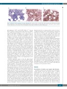
Figure 2. Immunophenotypic visualization of leukemic erythroblasts. Bone marrow sections of a patient with erythroid leukemia (acute erythroleukemia) stained with (A) Wright-Giemsa solution and antibodies against (B) glycophorin A and (C) E-cadherin. Note that virtually all leukemic erythroblasts co-express abundant amounts of glycophorin A and E-cadherin, thereby confirming the immature stage of maturation of leukemic (erythroid) cells.
glycophorin A, CD71, and CD105 (Table 2).53-56 Among these, CD105 is of special value for the detection of imma- ture erythropoietic progenitors in the bone marrow of patients with MDS. E-cadherin is also expressed specifi- cally on the surface of erythropoietic progenitor cells in human bone marrow.57 However, E-cadherin is not used routinely as an erythroid marker in flow cytometry stud- ies. Our faculty is of the opinion that CD105 and E-cad- herin should be validated further as routine diagnostic markers in erythroid disorders of the bone marrow. Another interesting aspect is that several cell surface anti- gens appear to be downregulated on erythroid progenitor cells in MDS patients.58 Among these antigens, the cox- sackie-adenovirus receptor (CAR) is a rather specific anti- gen: in fact, a decrease or lack of this receptor on erythroid progenitor cells is typically seen in patients with MDS and in other bone marrow neoplasms accompanied by marked dysplasia.58 Immunophenotypic studies are also of great importance to identify immature stages of erythropoiesis in patients with acute (myeloid/erythroid) leukemia in whom morphological and molecular studies alone are insufficient to establish a correct diagnosis (Figure 2 and Online Supplementary Figure S2). In these cases the use of immunohistochemical studies and multiparameter flow cytometry is essential.
Another important aspect is that the physiological development and maturation of red cells in the bone mar- row is regulated by the supporting microenvironment (consisting of macrophages and stromal cells) in so-called erythroid islands.59 These islands, also known as erythro- blastic islands, are considered to represent functional units and are detectable by conventional stains and specific immunological stains (see above) on bone marrow biop- sies. With age, the number of erythroid islands in the bone marrow decreases, while their size increases, which may suggest that the decrease in stem cell numbers and ery- throid progenitors during aging might be compensated by an increased proliferation of local erythroid progenitors (Figure 3).60 In patients with MDS, impaired formation of erythroid islands as well as structural abnormalities within these islands have also been described (Figure 3).60 In low- risk MDS, abnormal formation of erythroid islands may be the only histopathological change found in the bone marrow. Moreover, it has been described that erythroid island density correlates inversely with overall survival in patients with MDS.60 However, alterations of island con-
figuration and size (e.g. increased size) can also be detect- ed when an increase in red cell production is required in pathological conditions, such as in hemolytic anemia or acute blood loss. In patients with aplastic anemia and other bone marrow failure syndromes, only a few or no erythroid islands are found in pathological examinations, which we consider to be relevant diagnostically.
There are a number of established peripheral parameters through which red cell production can be assessed quanti- tatively. The most widely applied approach is to measure the numbers of reticulocytes in the peripheral blood. Another approach is to quantify the numbers of circulating erythroid progenitor cells, including multi-lineage colony- forming progenitors (CFU-GEMM) and burst-forming units (BFU-E). With this approach, clonal cytopenias (MDS) and aplastic anemia (in both conditions, CFU- GEMM and BFU-E are markedly reduced or absent) are separable from ‘reactive’ and ‘deficiency’ anemias in which BFU-E and CFU-GEMM are usually within normal ranges.61-63 Even in MDS patients with 5q-, who may devel- op thrombocytosis, erythropoiesis and BFU-E growth are usually suppressed. Factor (erythropoietin)-independent and erythropoietin-hyperresponsive growth of BFU-E is indicative of (almost diagnostic for) the presence of PV or a related myeloproliferative neoplasm (MPN).64,65 However, the colony-assay is rather tricky and its success depends on the experience of the team performing the test. Thus, although this assay is of practical value (and was previous- ly used as a criterion for PV), it is nowadays only employed routinely in a few specialized centers.
Anemia
Classification of anemia: novel aspects and etiologies
In general, anemia can be classified based on the regen- erative capacity of the bone marrow (hypo- or hyper- regenerative), the red cell volume (micro-, normo- or macrocytic), the etiology (bleeding, deficiency-induced, hemolytic, renal, inflammation-related, neoplastic, aplas- tic, others) and the dynamics with which anemia develops (acute, chronic).9-11 For each specific form of anemia, an extensive amount of published data has accumulated dur- ing the past few decades. A detailed description is beyond the scope of this article. There are also special variants of anemia that develop typically in the context of certain
haematologica | 2018; 103(10)
1597
