
Page 30 - 2018_10-Haematologica-web
P. 30
P. Valent et al.
induced by erythropoietin deprivation, HSP70 is exported from the nucleus and allows GATA-1 cleavage in ery- throid progenitor cells (Figure 1D).25-31 This model, in which the fate of erythroid precursors is determined by the localization of HSP70 in the nucleus, has been shown to be altered in ineffective erythropoiesis in various ane- mic conditions such as b-thalassemia, MDS,36 and congen- ital erythroblastopenia,37 and may provide a new concep- tual basis to improve anemia in these diseases.31
Apart from these established regulators of erythro- poiesis, several novel molecular players regulating ery- thropoiesis were discussed in the workshop. Among these are members of the BCL-2 family such as BID which may play a critical role in the regulation of mitochondrial depo- larization and caspase activation during erythroid differ- entiation and apoptosis (Figure 1C), various transcription factors, including STAT5A and STAT5B (Table 1), cytokines such as growth differentiating factor 11 (GDF11), and regulators of iron uptake and iron metabo- lism.33-48 GDF11, a ligand of activin receptor IIA (ActRIIA) that accumulates in erythroblasts in patients with b-tha- lassemia, has been implicated in the pathogenesis of ane- mia in these patients.42
It has also been described that polymeric immunoglob- ulin A (IgA) produced in the bone marrow may bind to the transferrin receptor-1 to sensitize erythroid cells to ery- thropoietin (Figure 1A).35 Consistent with this role as a regulator of erythropoiesis, the synthesis of polymeric IgA is increased during hypoxia. In pathological conditions, patients with IgA deficiency have higher levels of erythro- poietin and, conversely, patients with unexplained poly- cythemia associated with an excess of polymeric IgA syn- thesis have recently been reported.35 Finally, a number of regulators of red cell membrane stability have recently been identified. One of these regulators may be CDK6, a cell cycle regulator that has recently been shown to serve as a membrane stabilizer of red cells in mice.49
Diagnostic evaluation of erythropoiesis: established and novel markers
In most anemic patients the underlying etiology can be identified rapidly through the information gained from a thorough case history and detailed laboratory investiga- tions. A number of different etiologies can underlie ane- mia, including iron deficiency, chronic inflammation, hemolysis, renal disorders or vitamin B12 deficiency. In each case, it is important to follow the principle diagnostic algorithms, to establish the correct diagnosis and to treat the underlying disorder (for example a gastrointestinal dis- ease). In most of these cases, investigation of the bone marrow is not required. In other patients, however, the etiology remains uncertain after initial studies, so that detailed investigations of the bone marrow have to be per- formed. These investigations include a thorough cytologi- cal, histological and immuno-histochemical examination of the bone marrow, multi-parameter flow cytometry studies, a detailed examination of chromosomes (metaphases by conventional karyotyping and interphases by fluorescence in situ hybridization) and molecular stud- ies.9-11,50-52 Depending on blood counts and other parame- ters, fluorescence in situ hybridization studies are applied to screen for the presence of MDS-related abnormalities. Our faculty recommends that in each case morphological and immunophenotypic studies, including immunohisto- chemistry and flow cytometry, should be employed to
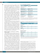
Table 1. Selected regulators of erythropoiesis.
Type of Regulator
Growth factors for multipotent and early erythropoietic progenitor cells
Later-acting erythropoietic
differentiation factors
Essential transcription factors
Important survival factors for
erythropoietic cells
Negative growth regulators of erythropoietic progenitor cells
Essential vitamins and
trace elements
Iron and proteins involved in iron distribution and iron metabolism
Major Examples
SCF, G-CSF, IL-3
EPO, TGF-beta, GDF11,
Activin A
GATA-1, STAT5A, STAT5B MCL-1, BCL-xL, HSP70*
Inhibin, TGF-beta, BID, FAS ligand, FAS, Caspases*
Vitamin B12, Folic acid,
Copper, others
Ferritin, Transferrin, Transferrin receptor (CD71) Ferroportin, Hepcidin
SCF: stem cell factor; G-CSF: granulocyte colony-stimulating factor; EPO: erythropoietin; TGF- beta: transforming growth factor beta; GDF11: growth differentiation factor 11, also known as bone morphogenetic protein 11; HSP70: heat shock protein 70. *HSP70 prevents caspase- induced cleavage of GATA1 in erythropoietic progenitor/precursor cells.
Table 2. Markers used to identify and quantify erythropoietic cells in the bone marrow.
Marker Ery-PC Ery
Specificity for erythropoietic cells
Flow cytometry
CD36/GP4
CD235a / Glycophorin A CD71 / TfR-1 E-Cadherin
CD105 / endoglin
Immunohistochemistry
CD235a / Glycophorin A CD236 / Glycophorin C** Hemoglobin A
CD71 / TfR-1 E-Cadherin*
CD105 / endoglin
++ + -
++ ++ ++ ++
++ ++ +/- ++ ++ ++
+++ ++ ++ -
+ +* +/- -
+++ ++ ++ ++ ++ ++ ++ +/- + +* +/- -
*Within the hematopoietic cell system, expression of E-cadherin is rather specific for red cells and their progenitors. **In immature erythroid leukemia, blast cells may stain negative for gly- cophorin A, but are still positive for glycophorin C and E-cadherin. Ery-PC: erythroid progenitor cells; Ery: erythrocytes; GP4: platelet plycoprotein-4; TfR-1: transferrin receptor-1.
define the percentage and stage of maturation of erythroid cells in bone marrow samples.52
For routine diagnostics, recommended immunohisto- chemical markers are glycophorin A, CD71 and E-cad- herin (Table 2). Additional immunohistochemical markers include glycophorin C and hemoglobin A. E-cadherin is of special value for the detection of immature erythropoietic progenitor cells in patients with MDS and those with ery- throleukemia (Figure 2). In contrast, hemoglobin A is pref- erentially expressed in more mature erythroblasts, and may therefore be helpful in distinguishing between more mature and more immature cells.
In flow cytometry studies, recommended markers are
1596
haematologica | 2018; 103(10)
