
Page 170 - 2018_10-Haematologica-web
P. 170
X. Wang et al.
stimulate the proliferation of HUVECs, suggesting that the pro-proliferative effect of TME5C was dependent on the specific amino acid sequence of this peptide (Figure 1C).
TME5C stimulates angiogenesis
The potential role of TME5C to stimulate angiogenesis was examined in vitro and in vivo. TME5C (500 nM) but not TME5A or TME5B stimulated vascular tube forma- tion of HUVECs and HHSECs by nearly 3-fold compared with control diluent-treated cells (Figure 2A-D). On the other hand, no remarkable proangiogenic effect was noted in the mutant form of TME5C (Figure 2E,F). Furthermore, in vivo angiogenesis assays with matrigel plugs revealed that TME5C (500 nM) stimulated angio- genesis in C57BL/6 mice (Figure 2G). In addition, the hemoglobin concentration was significantly increased in matrigel containing TME5C (Figure 2H) compared to matrigel containing control diluent. Consistent with the results of the proliferation assays, 30 nM TME5 produced an almost identical angiogenic effect with 500 nM TME5C (Figure 2A-D, G, H).
TME5C, but not TME5A or TME5B, blocks FK506-induced growth inhibition and apoptosis in endothelial cells
We examined whether TME5C counteracted the growth inhibition of HUVECs and HHSECs induced by the calcineurin inhibitor FK506. FK506 inhibited the pro- liferation of HUVECs and HHSECs by 80-90% and 70- 80%, respectively. This growth inhibition was signifi- cantly attenuated by the presence of TME5C in a dose- dependent manner up to 500 nM (Figure 3A). The highest dose of TME5C (1000 nM) was not as potent as 500 nM TME5C. Neither TME5A (500 nM) nor TME5B (500 nM) counteracted the effect of FK506 on proliferation of HUVECs and HHSECs (Figure 3A). We therefore chose 500 nM of TME5C for subsequent experiments. Importantly, the mutant form of TME5C lost the ability to block FK506-induced growth inhibition (Figure 3A). We next examined whether TME5C could block FK506- induced apoptosis in HUVECs and HHSECs. FK506 induced more than 40% of HUVECs and HHSECs to be apoptotic. Interestingly, when these cells were cultured in the presence of both FK506 (10 mg/mL) and TME5C (500 nM), the population of apoptotic cells significantly
AB
C
D
E
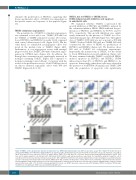
Figure 5. TME5C exerts cytoprotective function in murine ECs in a GPR15-dependent manner. (A). BrdU incorporation assays. WT or Gpr15 KO murine ECs were cultured with TME5C (500 nM) with or without FK506 (10 mg/ml) for 24 h. Proliferation was measured by BrdU incorporation assays. (B). Apoptosis assay. WT or Gpr15 KO murine ECs were exposed to FK506 (10 mg/ml) and/or TME5C (500 nM). After 36 h cells were harvested and stained with anti-annexin V and PI. FACS was used to analyze apoptotic cells. Annexin V+PI- and Annexin V+PI+ indicate early and late apoptosis, respectively. (C). Quantitative analysis of apoptotic cells in each group (n=3). (D, E). Vascular tube formation assays in vitro. WT or Gpr15 KO murine ECs were plated on growth factor-reduced matrigel-precoated 24-well plates (2.0x10⁴ cells/well) and incubated with control diluent, TME5C (500 nM), or VEGF (0.5 nM, positive control). After 8 h, the endothelial cell-derived tube-like structure was photographed using an inverted microscope. The tube length in three randomly chosen fields from each well was measured using NIH ImageJ software. Results represent the mean ± SD. *P<0.05. BrdU, bromodeoxyuridine; KO: knock out; GPR15: G-protein coupled receptor; WT: wild-type; FK506: tacrolimus; TM: thrombo- modulin; N.S.: not significant; VEGF: vascular endothelial growth factor.
1736
haematologica | 2018; 103(10)
