
Page 171 - 2018_10-Haematologica-web
P. 171
TME5C exerts cytoprotective and angiogenic functions
decreased (Figure 3B-E). By comparison, neither TME5A (500 nM) nor TME5B (500 nM) showed cytoprotective effects in HUVECs and HHSECs. In parallel with the induction of apoptosis, vascular permeability was pro- foundly induced in HUVECs after exposure to FK506 (10 mg/ml) for 12 h. Of note, FK506-induced vascular perme- ability was significantly attenuated in the presence of TME5C (500 nM), but not TME5A or TME5B (Figure 3F). Once more, a lower dose of TME5 (30 nM) produced a cytoprotective effect comparable to 500 nM TME5C (Figure 3).
TME5C upregulates p-ERK, p-AKT, p-p38, and Mcl-1 in endothelial cells
We examined whether TME5C acts through the intracel- lular signal transduction pathways in HUVECs and HHSECs. Western blot analysis with different antibodies against intracellular signal transduction pathways found that exposure of either HUVECs or HHSECs to TME5C (500 nM) but not TME5A (500 nM) or TME5B (500 nM) for 48 h significantly increased the levels of phospho (p)-ERK, p-AKT, p-p38, and Mcl-1 in these cells (Figure 4A-D).
GPR15 is indispensable for the effects of TME5C
Further experiments were carried out to test whether GPR15 also mediated the cytoprotective function of TME5C as it did for TME5. BrdU incorporation assays found that TME5C stimulated the proliferation of ECs isolated from WT C57BL/6 mice by nearly 1.5-fold com- pared with ECs treated with control diluent. In contrast, TME5C was not able to stimulate the proliferation of vas- cular ECs isolated from Gpr15 KO mice (Figure 5A). In
addition, TME5C significantly rescued the ECs isolated from WT C57BL/6 mice, but not Gpr15 KO mice, from FK506-induced growth inhibition and apoptosis (Figure 5A-C). Moreover, TME5C stimulated vascular tube for- mation in WT C57BL/6 murine ECs but not Gpr15 KO murine ECs (Figure 5D,E).
TME5C does not affect thrombin-mediated coagulation
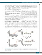
The fourth, fifth and sixth region of EGF-like domain of TM (TME456) binds thrombin and converts protein C to APC.32 The present study explored whether TME5C binds thrombin by measuring PT and APTT. rTM (500 nM) prolonged PT and APTT by approximately 200% and 165%, respectively. A much higher concentration of rTM (5000 nM) prolonged PT and APTT by more than 400% and 900%, respectively. Interestingly, TME5 (5000 nM) also prolonged PT and APTT by 137% and 225%, respectively. Of note, even the highest concentration of 5000 nM TME5C did not prolong either PT or APTT (Figure 6).
TME5C ameliorates SOS in a murine model
To induce SOS, we used a murine BMT model precon- ditioned with BU and CY (Figure 7A). BMT recipients treated with PBS showed a decrease in food intake, curled hairs, and abdominal distention (data not shown). Dark brown colored livers indicating congestion and massive ascites were noted in BMT recipients treated with PBS on day 7 after BMT (Figure 7B,C). In addition, liver enzymes, including ALT and AST, were significantly elevated in BMT recipients treated with PBS at day 7 (Figure 7D). On the other hand, these indicators were less significant in
AB
C
D
Figure 6. Effects of TME5C on PT and APTT. Plasma was obtained from a healthy volunteer and mixed with PT or APTT reagent with or without a series of TM frag- ments at various concentrations. (A, B). PT assays of TM fragments. (C, D). APTT assays of TM fragments. Experiments were performed three times. Results repre- sent the mean ± SD. *P<0.05 compared with control. PT: prothrombin time; APTT: activated partial thromboplastin time; TM: thrombomodulin; rTM: recombinant human soluble thrombomodulin.
haematologica | 2018; 103(10)
1737
