
Page 168 - 2018_10-Haematologica-web
P. 168
X. Wang et al.
TUNEL staining
Apoptosis of hepatocytes and sinusoidal ECs was assessed using an in situ cell death detection kit (Roche) according to the manufacturer’s protocol. Briefly, paraffin-embedded liver tissue sections were pretreated with dewaxation, rehydration, and proteinase K working solution, and subsequently the terminal deoxynucleotidyl transferase-mediated dUTP nick-end labeling (TUNEL) reaction mixture was added before adding the convert- er-POD. After the substrate solution was added, the slides were evaluated under a light microscope (×400).
ELISA
The plasma of mice was collected and analyzed with an enzyme-linked immunosorbent assay (ELISA) kit to measure the concentrations of TM, fibrinogen/fibrin degradation product (FDP), and plasminogen activator inhibitor-1 (PAI-1) according to the manufacturer’s protocol (Cloud-Clone Corp. Wuhan, China).
Statistical analysis
Statistical analyses were performed to assess the differences
A
B
C
DE
F
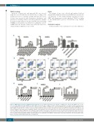
Figure 3. TME5C blocks apoptosis of FK506-treated endothelial cells. (A). BrdU incorporation assay. HUVECs or HHSECs were cultured with TME5C (25, 50, 250, 500, 1000 nM), TME5A (500 nM), TME5B (500 nM), TME5C mutant (500 nM), or TME5 (30 nM) in combination with FK506 (10 mg/ml) for 24 h. Proliferation was measured by BrdU incorporation assays (n=3). (B, C). Apoptosis assays. HUVECs or HHSECs were exposed to FK506 (10 mg/ml) with or without TME5A/B/C (500 nM) or TME5 (30 nM). After 36 h, cells were harvested, stained with anti-annexin V and PI, and subjected to FACS. Annexin V+PI- and Annexin V+PI+ indicate early and late apoptosis, respectively. (D, E). Quantitative analysis of the apoptotic cells in each group (n=3). (F). Vascular permeability assays. HUVEC monolayers were exposed to TME5 (30 nM) or TME5A/B/C (500 nM) with or without FK506 (10 mg/ml) for 12 h, and then fluorescein isothiocyanate-dextran was added. The fluores- cence of the plate well solution was measured to quantify the extent of permeability. Experiments were performed three times. Results represent the mean ± SD. *P<0.05. BrdU: bromodeoxyuridine; HUVECs: human umbilical vein endothelial cells; HHSECs: human hepatic sinusoidal endothelial cells; PI: propidium iodide. FK506: tacrolimus; TM: thrombomodulin; N.S.: not significant.
1734
haematologica | 2018; 103(10)
