
Page 167 - 2018_10-Haematologica-web
P. 167
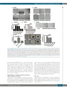
TME5C exerts cytoprotective and angiogenic functions
AB
CD
FG
E
H
Figure 2. TME5C stimulates angiogenesis in endothelial cells. (A, B, E). In vitro vascular tube formation assays. HUVECs or HHSECs were plated on growth factor- reduced matrigel-precoated 24-well plates (2.0x10⁴ cells/well) and incubated with control diluent, TME5 (30 nM), TME5A/B/C (500 nM), TME5C mutant (500 nM) or VEGF (0.5 nM, positive control). After 8 h, the endothelial cell-derived tube-like structure was photographed. (C, D, F). The tube length in three randomly chosen fields from each well was measured using NIH ImageJ software. (G, H). In vivo angiogenesis assays. Growth factor-reduced matrigel (0.3 ml, containing 40 U/ml heparin) with control diluent, TME5 (30 nM), TME5A/B/C (500 nM), or VEGF (0.5 nM, positive control) was subcutaneously injected into C57BL/6 mice near the abdominal midline (n=3 in each group). Four days later, mice were euthanized, and the matrigel plugs were dissected out and photographed. The matrigel plugs were homogenized in the presence of 1 ml distilled water and mixed with drabkin’s reagent. The hemoglobin levels were then measured using a microplate reader. Results represent the mean ± SD. *P<0.05. VEGF: vascular endothelial growth factor; HUVECs: human umbilical vein endothelial cells; HHSECs: human hepatic sinusoidal endothelial cells; TM: thrombomodulin; N.S.: not significant.
the mice intraperitoneally from day -7 to day -4 and from day - 3 to day -2, respectively. Two days later, the mice were intra- venously infused with bone marrow cells harvested from BALB/C mice (5×10⁶ per mouse). The day of BMT was set as day 0. Intraperitoneal administration of either TME5C (500 mg/kg) or vehicle PBS was initiated on day -7 and continued to day 13. Each agent was given to mice every other day. Blood was withdrawn, and plasma levels of aspartate aminotrans- ferase (AST) and alanine aminotransferase (ALT) were meas- ured on days 7, 14, and 20.
Hematoxylin-eosin (H&E), immunohistochemistry (IHC), and Masson staining
On days 7, 14, and 20 after BMT, some of the mice were sac- rificed. Livers were surgically removed and fixed with formalde- hyde solution. Specimens were dehydrated, waxed, and sliced into 4-mm thickness by an RM2126 microtome. After H&E staining, pathologic changes were evaluated under a light micro-
scope. Some liver slices were treated with 3% H2O2 and blocked with 1% bovine serum albumin (BSA). The slices were then incubated with primary pan-endothelial cell monoclonal anti- body (MECA-32, Novus Biologicals, Littleton, MA, USA) fol- lowed by incubation with biotinylated goat anti-rat secondary antibody and ABC HRP reagent. Color was developed with 3,3′- diaminobenzidine. Quantification of MECA-32-positive stained sinusoidal ECs was performed using NIH ImageJ software and expressed as the number of positive stained cells/analyzed area. Masson staining was carried out in accordance with the manu- facturer’s protocol (Sigma-Aldrich, Tokyo, Japan).
SOS score
Histological slices after H&E, IHC, or Masson staining were blindly evaluated according to the scoring system modified from that described by DeLeve et al.30 Based on the total score, the observed SOS was ranked as mild, moderate, or severe as previ- ously described.31
haematologica | 2018; 103(10)
1733
