
Page 110 - 2018_10-Haematologica-web
P. 110
R. Jimenez-P. et al.
CDCA7 silencing in these cells blocked or greatly impaired their tumor formation capacity (Figure 6) without substan- tially affecting the expression of the Ki67 proliferation marker (Online Supplementary Figure S10). These results strongly suggest that CDCA7 is critical for BL formation and that therapies aimed at inhibiting its expression or its activity might be of interest for BL patients. Since current therapy for BL patients affects not only tumor cells but also actively proliferating normal cells from these patients, we investigated whether CDCA7 silencing affected the prolif- eration of primary human diploid fibroblasts IMR-90. Transduction of these cells with lentivirus encoding sh-25 or sh-83 decreased CDCA7 mRNA and protein levels rela- tive to sh-Ctl-transduced cells (Online Supplementary Figures S11A-S11B) without affecting their proliferation rate (Online Supplementary Figure S11C).
CDCA7 expression is deregulated in lymphoid neoplasias and mediates their growth in vivo
Next, we assessed whether CDCA7 is deregulated not only in BL, but also in other types of cancer by comparing its expression in protein extracts of several lymphoid tumors. Cell lines of Diffuse Large B-cell Lymphoma (DLBCL), Follicular Lymphoma, Mantle Cell Lymphoma, uncharacterized non-Hodgkin Lymphoma, B-cell leukemia, and T-cell leukemia expressed markedly higher levels of CDCA7-2 than LCLs (Figure 7). In contrast, CDCA7-2 expression in myeloid leukemia cell lines was similar to that of LCLs (Figure 7, bottom panels).
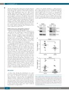
To investigate whether CDCA7 is an essential mediator of lymphomagenesis not only in BL, but also in other lym- phoid neoplasias, we silenced its expression in T-cell leukemia and DLBCL cells and analyzed their tumori- genicity in immunodeficient mice. Molt-4 T-cell leukemia and Toledo DLBCL cells expressed detectable levels of both CDCA7-1 and CDCA7-2 (Figure 8A) and their lentiviral transduction with sh-25 markedly inhibited the expression of both isoforms relative to cells transduced with sh-Ctl (Figure 8A). It should be noted that CDCA7-1 was not always detected in protein extracts from these cells. The capacity of these cells to form tumors was sub- sequently determined through their subcutaneous inocu- lation into immunodeficient mice. Molt-4 and Toledo cells transduced with sh-25 elicited formation of tumors signif- icantly smaller than those produced by cells transduced with sh-Ctl (Figures 8B-8C). Similar to BL cells, CDCA7 silencing in Toledo cells did not substantially affect cell cycle distribution (Online Supplementary Figure S12). Together, our results strongly suggest that CDCA7 is a key mediator of lymphoid malignant transformation.
Discussion
In this study, through the identification of a gene involved in anchorage-independent growth, we found a new target for therapeutic intervention in lymphoid tumors less prone to cause side effects. By comparing gene expression profiles of immortal, but non-malignant cells, with those of tumor cells from the same lineage, we have uncovered a gene, CDCA7, whose elevated protein levels in lymphoid tumor cells mediate their anchorage-indepen- dent growth and their tumorigenesis without participating in their growth under normal tissue culture conditions (liquid culture on a rigid surface).
CDCA7 was initially identified as a MYC-responsive gene19 whose expression can be transcriptionally induced also by E2F transcription factors22 and Notch.35 Although CDCA7 mRNA levels were shown to be upregulated in tumor samples relative to normal tissues, protein levels were not determined in these tumors.20 This is an impor- tant issue because the increase in mRNA expression does not necessarily imply an increase in protein content. Our results suggest that CDCA7 protein upregulation might be
A
B
C
Figure 8. CDCA7 mediates tumorigenesis in T-cell leukemia and DLBCL cells.
Toledo and Molt-4 cells were transduced with lentivirus encoding sh-Ctl or sh-25 and selected in the presence of puromycin >5 days. A. Representative CDCA7 immunoblot analysis of Toledo (n=3), and Molt-4 (n=3) cells expressing the indi- cated shRNAs. Tubulin is shown as loading control. B. Toledo and C. Molt-4 cells transduced with lentiviruses encoding the indicated shRNAs were inoculated subcutaneously in immunodeficient NOD-SCID mice. Tumors were extracted after 3 weeks. Circles and squares indicate the weight of individual tumors and horizontal bars indicate the mean (long bar) and s.e.m (short bar). **P<0.01 vs. sh-Ctl; paired samples t-test.
1676
haematologica | 2018; 103(10)
