
Page 96 - 2018_09-Mondo
P. 96
B.C. Ede et al.
AB
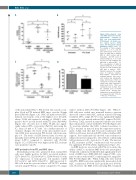
Figure 2. Mesenchymal stem cells (MSC) protect T-cell acute lymphoblastic leukemia (T- ALL) cells from parthenolide (PTL)-induced cell death, reac- tive oxygen species (ROS) stress and decreased reduced glutathione (rGSH) levels. (A) The viability of T-ALL samples (patients 1, 2, 5, 6, and 10) after 24 hours (h) of PTL treat- ment (10 μM) with or without MSC in direct contact and in transwells. (B) ROS levels in patient samples 2, 5, 6, 9, and 10 after 1 h PTL treatment (10
C
D μM) with or without MSC. (C) The concentration of rGSH in patient samples 2, 5, 6, 9, and 10 after 1 h PTL treatment (10 μM) with or without MSC. Symbols represent the average value in replicate samples. Each symbol represents an individual patient. Lines repre- sent median and interquartile range. (D) Increase in thiol con- centration in media from MSC culture after 24 h compared to blank media (n=6). Thiol con- centrations were derived from the standard curve of known cysteine levels. Results were analyzed by one-way ANOVA (A- C) or paired t-test (D). *P≤0.05,
**P≤0.01, ***P≤0.001.
of the antioxidant NAC to PTL treated cells caused a com- plete block in PTL induced ROS stress over the 90-min period (P≤0.02) (Figure 1A). NAC (15 mM) reversed PTL induced cytotoxicity, even at the highest dose (10 μM), where T-ALL cells retained a viability of 110±22% com- pared to 6±3% in cells treated with PTL alone (P≤0.0001) (Figure 1B). A lower dose of NAC, 30 μM, also signifi- cantly reduced PTL cytotoxicity (P≤0.01), increasing the IC50 from 2.3 μM to 5.7 μM. As a further measure of oxidative changes, the levels of the anti-oxidative mole- cule rGSH were measured in PTL treated cells from sam- ples 1-9. The levels of rGSH detected after 1 h were sig- nificantly lower in PTL treated cells with a median of 12% (range 0-58%) compared to untreated cells (P=0.02) (Figure 1C). rGSH levels were higher in the PTL resistant cases (patients 3 and 9).
MSC protection from PTL and ROS stress
Mesenchymal stem cells generated from normal BM cells were highly positive for MSC associated markers; CD73 (99±0.3%), CD105 (90±3%), CD90 (99±0.2%) with low expression of hemopoietic cell markers CD45 (0.3%±0.2%) and CD34 (0.6±0.5%). MSC protected T- ALL cells from the cytotoxic effects of PTL (78% median survival, range 57-84) in co-culture compared to 8% (range
2-26%) without MSC (P=0.002) (Figure 2A). When T- ALL cells were seeded onto transwell inserts to prevent direct cell contact with MSC, median survival following treatment (58%, range 20-71%) was significantly higher compared to cells treated without MSC support (P=0.03). However, T-ALL survival in transwell cultures was lower than cells in direct contact with MSC (P=0.02) (Figure 2A). To ascertain whether this MSC conferred resistance to PTL is retained on removal from the supportive environ- ment, T-ALL cells that had been pre-conditioned with MSC for 24 h were treated with PTL without further MSC support. Pre-conditioned cells showed some evidence for enhanced survival compared to cells without conditioning with an increase in IC50 from 2.6 to 3.3 μM. The increase in PTL resistance in MSC conditioned cells was modest but significant (P=0.04) (Online Supplementary Figure S3).
As ROS levels are associated with PTL cytotoxicity, the ability of MSC to modulate ROS levels was investigated. T-ALL cells conditioned with MSC had significantly lower levels of ROS stress, (ROS MFI 234, range 67-380), com- pared to those without MSC conditioning (313, range 156- 506; P=0.02) (Figure 2B). When T-ALL cells were treated with PTL, ROS levels were significantly increased in both MSC conditioned (P=0.04) and unconditioned (P=0.01) cells. However, the ROS levels following PTL treatment
1496
haematologica | 2018; 103(9)
