
Page 94 - 2018_09-Mondo
P. 94
B.C. Ede et al.
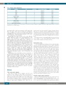
Table 1. Patients’ sample characteristics.
Patient N.
1
2 3 4 5 6 7 8 9
10
Karyotype Sex
46XY M
t(1;14) M 46XY M t(8;14) M t(11;14), add(17) M Runx1 rearr M 46XY M add (4), add (9) M del (4), del (9) M
add (7), add (9), add (14) M
Age (years)
7
3
17
3
2
3
6
14
9
4
Disease status at biopsy
Diagnosis
Diagnosis
Diagnosis
Diagnosis
Relapse
Diagnosis
Diagnosis
Diagnosis
Diagnosis
Diagnosis
MRD risk statusa
Risk
Risk
High
Low
High
N/A
Low
High
Low
Low
All patients treated on UKALL2011 protocol, except patients 3, 5 and 8 who were treated on UKALL 2003. aMinimal residual disease (MRD) risk status at Day 29, on respective treatment protocols. N: number; M: male.
on normal cells at the doses required to kill cancer cells. PTL can target cancer cells via numerous mechanisms, such as inhibition of nuclear factor (κ)B, p53 activation and ROS stress.6,7 However, the mechanism of PTL toxici- ty to T-ALL has not been defined.
Parthenolide has been shown to be very effective against childhood T-ALL in vivo, with elimination of the disease and restoration of murine hemopoiesis in NOD.Cg-PrkdcscidIl2rtm1Wjl/SzJ (NSG) mice.8 However, in mice engrafted with different leukemia initiating cell pop- ulations from 2 of 9 T-ALL cases, disease progression was delayed rather than eliminated, indicating variable sensi- tivity of certain subpopulations to PTL. Reasons for the differences in sensitivity may be due to the effect of the in vivo microenvironment. Bone marrow (BM) stromal cells release cysteine for uptake by chronic lymphocytic leukemia (CLL) cells, driving anti-oxidative glutathione synthesis, which provides protection against ROS gener- ating chemotherapeutic agents, such as fludarabine and oxiplatin.9
Mesenchymal stem cells (MSC) are key constituents of the BM microenvironment and have been shown to enhance protection against certain drugs in T-ALL cell lines10 and primary samples from patients with B-ALL, acute myeloid leukemia (AML) and CLL.9,11-13 Co-culture of T-ALL cell lines with MSC enhanced resistance to the anthracycline idarubicin.10 However, the role of ROS in stromal cell mediated protection in childhood ALL has not been reported. As we had previously reported resistance to PTL in T-ALL cases, in this study the cytotoxic and ROS inducing effects of the drug on primary T-ALL cells in the presence of MSC were examined to increase our under- standing of PTL resistance.
Methods
T-ALL and normal samples
Bone marrow samples from 10 children, aged 2-17 years (median 5 years), diagnosed with T-ALL at presentation or relapse were collected with informed consent and approval of University Hospitals Bristol NHS Trust and London Brent Research Ethics Committee (Table 1). Mononuclear cells (MNC) were separated via density gradient centrifugation using Ficoll- Hypaque (Sigma-Aldrich, Gillingham, UK). MNC were sus-
pended in 90% fetal calf serum (FCS, Thermo Scientific, Paisley, UK) and 10% dimethyl sulfoxide (DMSO, Origen Biomedical, Solihull, UK) and stored in liquid nitrogen prior to use. Samples from patients with a range of karyotypic abnormalities, diagnos- tic age and minimal residual disease (MRD) status were investi- gated.
Bone marrow from a consenting healthy donor was used as a source of MSC. See the Online Supplementary Appendix for full details of MSC expansion and characterization.
Cytotoxicity assays
T-cell acute lymphoblastic leukemia cells were plated in dupli- cate (for each drug concentration tested) at 1.2x10⁵ cells/mL in RPMI 1640 medium (Sigma-Aldrich) containing 20% FCS, 1% L- glut and 1% Pen/Strep, hereafter referred to as suspension medi- um. Drugs used for assays were: PTL (Enzo Life Sciences, Exeter, UK) at 1-10 μM, N-acetyl cysteine (NAC, Sigma-Aldrich) at 15 mM and 30 μM, and sulfasalazine (SSZ, Sigma-Aldrich) at 300 μM, all prepared in suspension medium. For co-culture experiments, 5x10⁴ MSC/mL were seeded per well and left to adhere for 24 hours (h). MSC medium was removed and replaced with suspension media containing 1x10⁵ T-ALL cells/mL. T-ALL cells were left to settle for 1 h and then treated with 10 μM PTL with or without 300 μM SSZ, for 24 h. After treatment, non-adherent cells were removed and stained with annexin-V conjugated to fluorescein isothiocyante (Miltenyi Biotec) for 10 minutes (min). Cells were washed and stained with propidium iodide (PI, Miltenyi Biotec) prior to flow cyto- metric analysis. See Online Supplementary Figure S1 for details.
For transwell separation experiments, 1x10⁵ MSC/mL were seeded per well in MSC media and left to adhere for 24 h. MSC medium was removed and replaced with suspension medium. T-ALL cells were seeded at 2x10⁵ cells/mL onto Costar Transwell inserts (0.4 μM pore size, Corning Life Sciences, Ewloe, UK) above the adherent MSC. T-ALL cells were left to settle for 1 h and then treated with PTL with or without SSZ for 24 h. After treatment, cells in transwell inserts were removed and viability was assessed by flow cytometry, as above.
Reactive oxygen species detection
Cells were treated with 5 μM of the redox sensitive probe 5- (and-6)-chloromethyl-2',7'-dichlorodihydrofluorescein di- acetate (CM-H2DCFDA, Thermo Fisher Scientific) for 30 min at 37°C. Cells were treated with PTL, then immediately analyzed
1494
haematologica | 2018; 103(9)
