
Page 77 - 2018_09-Mondo
P. 77
Inhibition of DHODH induces differentiation in AML
AML cell differentiation. We measured the level of expression four myeloid differentiation markers, CD14, CD11b, CD33 and CD34, by flow cytometry. Of note, we found that isobavachalcone increased the expression of CD14 and CD11b (Figure 4A,B), but had no effect on the expression of CD33 and CD34 (Online Supplementary Figure S7A) in HL60 cells. As DHODH is involved in the
AF
B
CG
intracellular synthesis of uridine,29 we observed that isobavachalcone treatment lead to the depletion of uri- dine in HL60 cells (Online Supplementary Figure S8A). The uridine rescue experiment demonstrates that the endoge- nous cellular pyrimidine deficiency induced by DHODH inhibition is crucial for the differentiation of AML cells (Figure 4A,B and Online Supplementary Figure S7B).
D
E
H
I
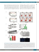
Figure 3. Isobavachalcone shows anti-proliferative activity against acute myeloid leukemia cells. (A) HL60 and THP1 cells were treated with increasing concentra- tions of isobavachalcone for 48 h, and cell viability was measured by MTS assay. (B) The time-response curve of 30 μM isobavachalcone on cell viability of HL60 and THP1 cells. (C) Recombinant DHODH protein or HL60 cell lysate was incubated with control or isobavachalcone-conjugated Sepharose 4B beads. Proteins bound to the beads were analyzed by western blot. (D) Cellular thermal shift assay shows that isobavachalcone stabilizes and targets DHODH in intact HL60 cells. Cells were incubated with isobavachalcone for 12 h and the assay was performed. (E) The IC50 value of isobavachalcone against DHODH-knockout HL60 cells. (F) HL60 cells were treated with isobavachalcone at the indicated concentrations for 72 h. Cell apoptosis was detected by flow cytometry using staining with annexin V, fluo- rescein isothiocyanate (FITC) and propidium iodine (PI). (G) The quantitative data of cell apoptosis in (F). (H) Changes in apoptosis-related proteins after treatment with isobavachalcone or uridine for 72 h. (I) Representative images of Hoechst 33258-stained cells were analyzed by fluorescence microscopy in HL60 cells treated with 30 μM isobavachalcone for 48 h. Red arrows indicate apoptotic cells. IBC: isobavachalocone.
haematologica | 2018; 103(9)
1477
